5Y04

Crystal Structure of the complex between the vinculin D1 domain and alphaE-catenin


Experimental Data Snapshot

  • Method: X-RAY DIFFRACTION
  • Resolution: 2.85 Å
  • R-Value Free: 0.281 
  • R-Value Work: 0.247 
  • R-Value Observed: 0.248 

wwPDB Validation   3D Report Full Report


This is version 1.2 of the entry. See complete history


Literature

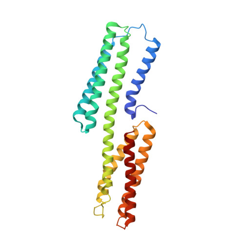
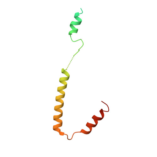
The force-sensing device region of alpha-catenin is an intrinsically disordered segment in the absence of intramolecular stabilization of the autoinhibitory form

Hirano, Y.Amano, Y.Yonemura, S.Hakoshima, T.

(2018) Genes Cells 23: 370-385

  • DOI: https://doi.org/10.1111/gtc.12578
  • Primary Citation of Related Structures:  
    5XFL, 5Y04

  • PubMed Abstract: 

    Mechanotransduction by α-catenin facilitates the force-dependent development of adherens junctions (AJs) by recruiting vinculin to reinforce actin anchoring of AJs. The α-catenin mechanotransducing action is facilitated by its force-sensing device region that autoinhibits the vinculin-binding site 1 (VBS1). Here, we report the high-resolution structure of the force-sensing device region of α-catenin, which shows the autoinhibited form comprised of helix bundles E, F and G. The cryptic VBS1 is embedded into helix bundle E stabilized by direct interactions with the autoinhibitory region forming helix bundles F and G. Our molecular dissection study showed that helix bundles F and G are stable in solution in each isolated form, whereas helix bundle E that contains VBS1 is unstable and intrinsically disordered in solution in the isolated form. We successfully identified key residues mediating the autoinhibition and produced mutated α-catenins that display variable force sensitivity and autoinhibition. Using these mutants, we demonstrate both in vitro and in vivo that, in the absence of this stabilization, the helix bundle containing VBS1 would adopt an unfolded form, thus exposing VBS for vinculin binding. We provide evidence for importance of mechanotransduction with the intrinsic force sensitivity for vinculin recruitment to adherens junctions of epithelial cell sheets with mutated α-catenins.


  • Organizational Affiliation

    Structural Biology Laboratory, Nara Institute of Science and Technology, Ikoma, Nara, Japan.


Macromolecules
Find similar proteins by:  (by identity cutoff)  |  3D Structure
Entity ID: 1
MoleculeChains Sequence LengthOrganismDetailsImage
Vinculin252Mus musculusMutation(s): 0 
Gene Names: Vcl
UniProt
Find proteins for Q64727 (Mus musculus)
Explore Q64727 
Go to UniProtKB:  Q64727
Entity Groups  
Sequence Clusters30% Identity50% Identity70% Identity90% Identity95% Identity100% Identity
UniProt GroupQ64727
Sequence Annotations
Expand
  • Reference Sequence
Find similar proteins by:  (by identity cutoff)  |  3D Structure
Entity ID: 2
MoleculeChains Sequence LengthOrganismDetailsImage
Catenin alpha-1102Mus musculusMutation(s): 0 
Gene Names: Ctnna1Catna1
UniProt
Find proteins for P26231 (Mus musculus)
Explore P26231 
Go to UniProtKB:  P26231
Entity Groups  
Sequence Clusters30% Identity50% Identity70% Identity90% Identity95% Identity100% Identity
UniProt GroupP26231
Sequence Annotations
Expand
  • Reference Sequence
Experimental Data & Validation

Experimental Data

  • Method: X-RAY DIFFRACTION
  • Resolution: 2.85 Å
  • R-Value Free: 0.281 
  • R-Value Work: 0.247 
  • R-Value Observed: 0.248 
  • Space Group: P 21 21 2
Unit Cell:
Length ( Å )Angle ( ˚ )
a = 54.807α = 90
b = 73.825β = 90
c = 111.485γ = 90
Software Package:
Software NamePurpose
HKL-2000data reduction
HKL-2000data scaling
SHARPphasing
REFMACrefinement

Structure Validation

View Full Validation Report


Entry History 

Deposition Data

Revision History  (Full details and data files)

  • Version 1.0: 2018-03-28
    Type: Initial release
  • Version 1.1: 2018-05-23
    Changes: Data collection, Database references
  • Version 1.2: 2024-03-27
    Changes: Data collection, Database references