The trimeric solution structure and fucose-binding mechanism of the core fucosylation-specific lectin PhoSL.
Yamasaki, K., Yamasaki, T., Tateno, H.(2018) Sci Rep 8: 7740-7740
- PubMed: 29773815 Search on PubMedSearch on PubMed Central
- DOI: https://doi.org/10.1038/s41598-018-25630-2
- Primary Citation Related Structures:
5XZK - PubMed Abstract:
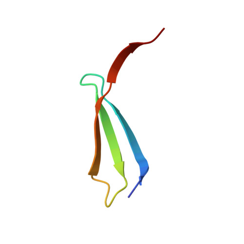
The core α1-6 fucosylation-specific lectin from a mushroom Pholiota squarrosa (PhoSL) is a potential tool for precise diagnosis of cancers. This lectin consists of only 40 amino acids and can be chemically synthesized. We showed here that a synthesized PhoSL peptide formed a trimer by gel filtration and chemical cross-linking assays, and determined a structure of the PhoSL trimer by NMR. The structure possesses a β-prism motif with a three-fold rotational symmetry, where three antiparallel β-sheets are tightly connected by swapping of β-strands. A triad of Trp residues comprises the structural core, forming NH-π electrostatic interactions among the indole rings. NMR analysis with an excess amount of fucose revealed the structural basis for the molecular recognition. Namely, fucose deeply enters a pocket formed at a junction of β-sheet edges, with the methyl group placed at the bottom. It forms a number of hydrophobic and hydrogen-bonding interactions with PhoSL residues. In spite of partial similarities to the structures of other functionally related lectins, the arrangement of the antiparallel β-sheets in the PhoSL trimer is novel as a structural scaffold, and thus defines a novel type of lectin structure.
- Biomedical Research Institute, National Institute of Advanced Industrial Science and Technology (AIST), Tsukuba, 305-8566, Japan. k-yamasaki@aist.go.jp.
Organizational Affiliation: