Substrate-bound structures of a ketoreductase from amphotericin modular polyketide synthase.
Liu, C., Yuan, M., Xu, X., Wang, L., Keatinge-Clay, A.T., Deng, Z., Lin, S., Zheng, J.(2018) J Struct Biol 203: 135-141
- PubMed: 29626512 Search on PubMedSearch on PubMed Central
- DOI: https://doi.org/10.1016/j.jsb.2018.04.001
- Primary Citation Related Structures:
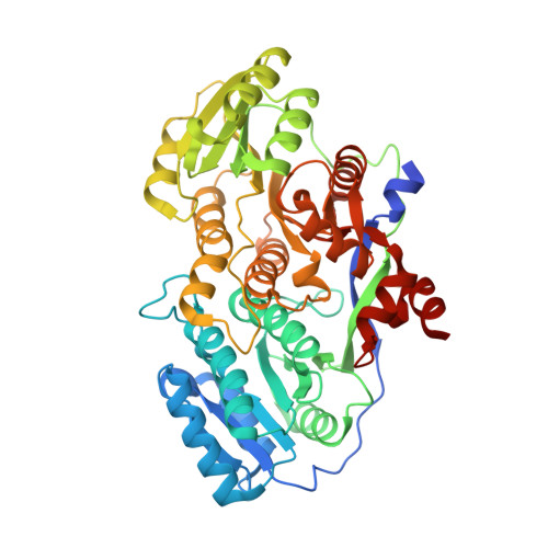
5XWV, 5XWW - PubMed Abstract:
Ketoreductase (KR) domains of modular polyketide synthases (PKSs) control the stereochemistry of C2 methyl and C3 hydroxyl substituents of polyketide intermediates. To understand the molecular basis of stereocontrol exerted by KRs, the crystal structure of a KR from the second module of the amphotericin PKS (AmpKR2) complexed with NADP + and 2-methyl-3-oxopentanoyl-pantetheine was solved. This first ternary structure provides direct evidence to the hypothesis that a substrate enters into the active site of an A-type KR from the side opposite the coenzyme to generate an L-hydroxyl substituent. A comparison with the ternary complex of a G355T/Q364H mutant sheds light on the structural basis for stereospecificity toward the substrate C2 methyl substituent. Functional assays suggest the pantetheine handle shows obvious influence on the catalytic efficiency and the stereochemical outcome. Together, these findings extend our current understanding of the stereochemical control of PKS KR domains.
- State Key Laboratory of Microbial Metabolism, School of Life Sciences and Biotechnology, Shanghai Jiao Tong University, Shanghai 200240, China.
Organizational Affiliation: