


Crystal structures of acyl carrier protein synthases (AcpS) from three Gram-negative bacteria
Liao, Y.P., Wang, D.L., Yin, D.P., Zhang, Q.Y., Wang, Y.M., Wang, D.Q., Zhu, H.X., Chen, S.To be published.
Experimental Data Snapshot
Starting Model: experimental
View more details
Entity ID: 1 | |||||
|---|---|---|---|---|---|
| Molecule | Chains | Sequence Length | Organism | Details | Image |
| Holo-[acyl-carrier-protein] synthase | 126 | Escherichia coli K-12 | Mutation(s): 1 Gene Names: acpS, dpj, b2563, JW2547 EC: 2.7.8.7 | 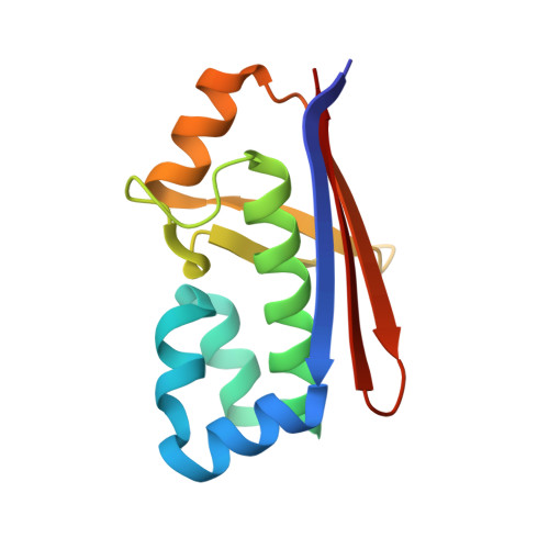 | |
UniProt | |||||
Entity Groups | |||||
| Sequence Clusters | 30% Identity50% Identity70% Identity90% Identity95% Identity100% Identity | ||||
| UniProt Group | P24224 | ||||
Sequence AnnotationsExpand | |||||
Reference Sequence | |||||
| Ligands 3 Unique | |||||
|---|---|---|---|---|---|
| ID | Chains | Name / Formula / InChI Key | 2D Diagram | 3D Interactions | |
| COA Download:Ideal Coordinates CCD File | H [auth B], J [auth C] | COENZYME A C21 H36 N7 O16 P3 S RGJOEKWQDUBAIZ-IBOSZNHHSA-N |  | ||
| GOL Download:Ideal Coordinates CCD File | G [auth A] | GLYCEROL C3 H8 O3 PEDCQBHIVMGVHV-UHFFFAOYSA-N |  | ||
| CL Download:Ideal Coordinates CCD File | D [auth A], E [auth A], F [auth A], I [auth B], K [auth C] | CHLORIDE ION Cl VEXZGXHMUGYJMC-UHFFFAOYSA-M |  | ||
| Length ( Å ) | Angle ( ˚ ) |
|---|---|
| a = 97.8 | α = 90 |
| b = 97.8 | β = 90 |
| c = 95.38 | γ = 90 |
| Software Name | Purpose |
|---|---|
| REFMAC | refinement |
| iMOSFLM | data reduction |
| SCALA | data scaling |
| PHASER | phasing |


