5XSY | pdb_00005xsy

Structure of the Nav1.4-beta1 complex from electric eel


Experimental Data Snapshot

  • Method: ELECTRON MICROSCOPY
  • Resolution: 4.00 Å
  • Aggregation State: PARTICLE 
  • Reconstruction Method: SINGLE PARTICLE 

wwPDB Validation 3D Report Full Report

Validation slider image for 5XSY

This is version 2.2 of the entry. See complete history

Literature

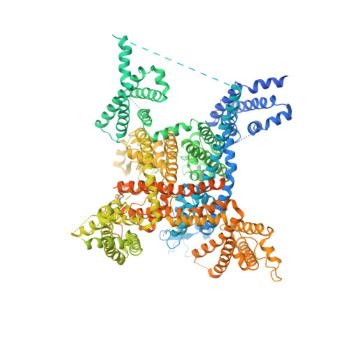
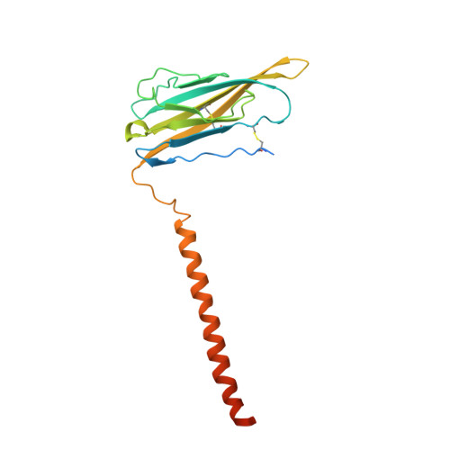
Structure of the Nav1.4-beta 1 Complex from Electric Eel.

Yan, Z.Zhou, Q.Wang, L.Wu, J.Zhao, Y.Huang, G.Peng, W.Shen, H.Lei, J.Yan, N.

(2017) Cell 170: 470-482.e11

  • DOI: https://doi.org/10.1016/j.cell.2017.06.039
  • Primary Citation Related Structures: 
    5XSY

  • PubMed Abstract: 

    Voltage-gated sodium (Na v ) channels initiate and propagate action potentials. Here, we present the cryo-EM structure of EeNa v 1.4, the Na v channel from electric eel, in complex with the β1 subunit at 4.0 Å resolution. The immunoglobulin domain of β1 docks onto the extracellular L5 I and L6 IV loops of EeNa v 1.4 via extensive polar interactions, and the single transmembrane helix interacts with the third voltage-sensing domain (VSD III ). The VSDs exhibit "up" conformations, while the intracellular gate of the pore domain is kept open by a digitonin-like molecule. Structural comparison with closed Na v PaS shows that the outward transfer of gating charges is coupled to the iris-like pore domain dilation through intricate force transmissions involving multiple channel segments. The IFM fast inactivation motif on the III-IV linker is plugged into the corner enclosed by the outer S4-S5 and inner S6 segments in repeats III and IV, suggesting a potential allosteric blocking mechanism for fast inactivation.


  • Organizational Affiliation
    • State Key Laboratory of Membrane Biology, Tsinghua-Peking Joint Center for Life Sciences, School of Life Sciences and School of Medicine, Tsinghua University, Beijing, China; Beijing Advanced Innovation Center for Structural Biology, School of Life Sciences, Tsinghua University, Beijing, China.

Macromolecule Content 

  • Total Structure Weight: 235.27 kDa 
  • Atom Count: 10,634 
  • Modeled Residue Count: 1,312 
  • Deposited Residue Count: 2,029 
  • Unique protein chains: 2

Macromolecules

Find similar proteins by:|  3D Structure
Entity ID: 1
MoleculeChains  Sequence LengthOrganismDetailsImage
Sodium channel protein1,820Electrophorus electricusMutation(s): 0 
Membrane Entity: Yes 
UniProt
Find proteins for P02719 (Electrophorus electricus)
Explore P02719 
Go to UniProtKB:  P02719
Entity Groups
Sequence Clusters30% Identity50% Identity70% Identity90% Identity95% Identity100% Identity
UniProt GroupP02719
Glycosylation
Glycosylation Sites: 3
Sequence Annotations
Expand
Reference Sequence
Find similar proteins by:|  3D Structure
Entity ID: 2
MoleculeChains  Sequence LengthOrganismDetailsImage
Voltage-gated sodium channel beta subunit 1209Electrophorus electricusMutation(s): 0 
Membrane Entity: Yes 
UniProt
Find proteins for A0A1L3MZ94 (Electrophorus electricus)
Explore A0A1L3MZ94 
Go to UniProtKB:  A0A1L3MZ94
Entity Groups
Sequence Clusters30% Identity50% Identity70% Identity90% Identity95% Identity100% Identity
UniProt GroupA0A1L3MZ94
Glycosylation
Glycosylation Sites: 2
Sequence Annotations
Expand
Reference Sequence

Oligosaccharides

Help  
Entity ID: 3
MoleculeChains Length2D Diagram GlycosylationD Interactions
beta-D-mannopyranose-(1-3)-[beta-D-mannopyranose-(1-6)]beta-D-mannopyranose-(1-4)-2-acetamido-2-deoxy-beta-D-glucopyranose-(1-4)-2-acetamido-2-deoxy-beta-D-glucopyranose
C
5N-Glycosylation
Glycosylation Resources
GlyTouCan: G89225LT
GlyCosmos: G89225LT
GlyGen: G89225LT
Entity ID: 4
MoleculeChains Length2D Diagram GlycosylationD Interactions
beta-D-mannopyranose-(1-4)-2-acetamido-2-deoxy-beta-D-glucopyranose-(1-4)-2-acetamido-2-deoxy-beta-D-glucopyranose
D, E, F
3N-Glycosylation
Glycosylation Resources
GlyTouCan: G15407YE
GlyCosmos: G15407YE
GlyGen: G15407YE
Entity ID: 5
MoleculeChains Length2D Diagram GlycosylationD Interactions
2-acetamido-2-deoxy-beta-D-glucopyranose-(1-4)-2-acetamido-2-deoxy-beta-D-glucopyranose
G
2N-Glycosylation
Glycosylation Resources
GlyTouCan: G42666HT
GlyCosmos: G42666HT
GlyGen: G42666HT

Experimental Data & Validation

Experimental Data

  • Method: ELECTRON MICROSCOPY
  • Resolution: 4.00 Å
  • Aggregation State: PARTICLE 
  • Reconstruction Method: SINGLE PARTICLE 
EM Software:
TaskSoftware PackageVersion
MODEL REFINEMENTPHENIX

Structure Validation

View Full Validation Report


Entry History 

& Funding Information

Deposition Data


Funding OrganizationLocationGrant Number
Ministry of Science and Technology of ChinaChina2015CB910101, 2016YFA0500402, 2014ZX09507003-006
National Natural Science Foundation of ChinaChinaprojects 31621092, 31630017, 31611130036

Revision History  (Full details and data files)

  • Version 1.0: 2017-08-09
    Type: Initial release
  • Version 1.1: 2018-10-24
    Changes: Data collection, Other, Refinement description
  • Version 2.0: 2020-07-29
    Type: Remediation
    Reason: Carbohydrate remediation
    Changes: Atomic model, Data collection, Derived calculations, Structure summary
  • Version 2.1: 2024-10-30
    Changes: Data collection, Database references, Derived calculations, Structure summary
  • Version 2.2: 2025-07-02
    Changes: Data collection