5XQL

Crystal structure of a Pseudomonas aeruginosa transcriptional regulator


Experimental Data Snapshot

  • Method: X-RAY DIFFRACTION
  • Resolution: 2.49 Å
  • R-Value Free: 0.275 
  • R-Value Work: 0.232 
  • R-Value Observed: 0.234 

wwPDB Validation   3D Report Full Report


Ligand Structure Quality Assessment 


This is version 1.3 of the entry. See complete history


Literature

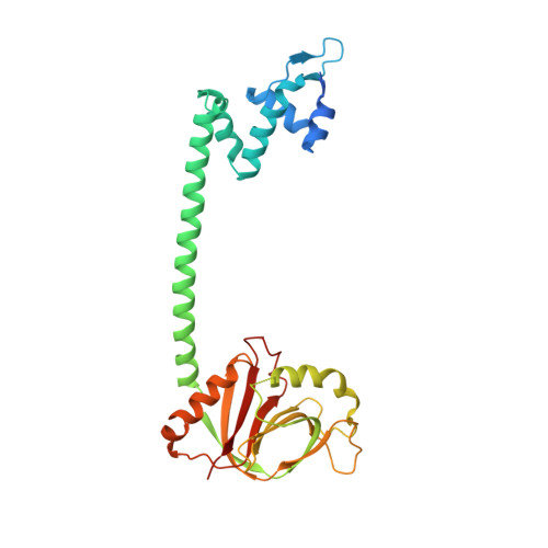
Crystal structure of BrlR with c-di-GMP

Raju, H.Sharma, R.

(2017) Biochem Biophys Res Commun 490: 260-264

  • DOI: https://doi.org/10.1016/j.bbrc.2017.06.033
  • Primary Citation of Related Structures:  
    5XQL

  • PubMed Abstract: 

    The transcriptional regulator BrlR is a member of the MerR family of multidrug transport activators in Pseudomonas aeruginosa. Recent study indicates that BrlR is a novel 3',5'-cyclic diguanylic acid (c-di-GMP) receptor and can be activated by c-di-GMP. To gain insight into BrlR function, we determined the structure of BrlR with c-di-GMP complex structure to 2.5 Å. The structure and size exclusion chromatography (SEC) data revealed BrlR forms a tetramer and each BrlR protomer consists of three parts, DNA-binding domain, a coiled-coil region and GyrI-like domain. There are two different c-di-GMP binding sites located mainly at the DNA binding domain of each BrlR protomer and do not overlap with the GyrI-like domain. The drug-binding pocket in GyrI-like domain is much conserved indicating it can also bind flat-shaped molecules like other multidrug resistance (MDR) proteins.


  • Organizational Affiliation

    Department of Biophysics, Molecular Biology and Bioinformatics, University of Calcutta, Kolkata, 700009, West Bengal, India.


Macromolecules
Find similar proteins by:  (by identity cutoff)  |  3D Structure
Entity ID: 1
MoleculeChains Sequence LengthOrganismDetailsImage
Multidrug-efflux transporter 1 regulator291Pseudomonas aeruginosaMutation(s): 0 
Gene Names: bmrRAO964_25340AOY09_00728B0B20_02615PAERUG_E15_London_28_01_14_00871PAERUG_P32_London_17_VIM_2_10_11_01154
UniProt
Find proteins for Q9HUT5 (Pseudomonas aeruginosa (strain ATCC 15692 / DSM 22644 / CIP 104116 / JCM 14847 / LMG 12228 / 1C / PRS 101 / PAO1))
Explore Q9HUT5 
Go to UniProtKB:  Q9HUT5
Entity Groups  
Sequence Clusters30% Identity50% Identity70% Identity90% Identity95% Identity100% Identity
UniProt GroupQ9HUT5
Sequence Annotations
Expand
  • Reference Sequence
Small Molecules
Ligands 1 Unique
IDChains Name / Formula / InChI Key2D Diagram3D Interactions
C2E
Query on C2E

Download Ideal Coordinates CCD File 
B [auth A],
C [auth A]
9,9'-[(2R,3R,3aS,5S,7aR,9R,10R,10aS,12S,14aR)-3,5,10,12-tetrahydroxy-5,12-dioxidooctahydro-2H,7H-difuro[3,2-d:3',2'-j][1,3,7,9,2,8]tetraoxadiphosphacyclododecine-2,9-diyl]bis(2-amino-1,9-dihydro-6H-purin-6-one)
C20 H24 N10 O14 P2
PKFDLKSEZWEFGL-MHARETSRSA-N
Experimental Data & Validation

Experimental Data

  • Method: X-RAY DIFFRACTION
  • Resolution: 2.49 Å
  • R-Value Free: 0.275 
  • R-Value Work: 0.232 
  • R-Value Observed: 0.234 
  • Space Group: I 4 2 2
Unit Cell:
Length ( Å )Angle ( ˚ )
a = 135.944α = 90
b = 135.944β = 90
c = 95.127γ = 90
Software Package:
Software NamePurpose
PHENIXrefinement
iMOSFLMdata reduction
Aimlessdata scaling
PHASERphasing

Structure Validation

View Full Validation Report


Ligand Structure Quality Assessment 


Entry History 

Deposition Data

Revision History  (Full details and data files)

  • Version 1.0: 2017-06-28
    Type: Initial release
  • Version 1.1: 2017-07-05
    Changes: Database references
  • Version 1.2: 2017-08-02
    Changes: Database references
  • Version 1.3: 2024-03-27
    Changes: Data collection, Database references, Structure summary