5XLK | pdb_00005xlk

Crystal structure of the flagellar cap protein FliD D2-D3 domains from Serratia marcescens in Space group I422


Experimental Data Snapshot

  • Method: X-RAY DIFFRACTION
  • Resolution: 3.05 Å
  • R-Value Free: 
    0.287 (Depositor), 0.286 (DCC) 
  • R-Value Work: 
    0.238 (Depositor), 0.238 (DCC) 
  • R-Value Observed: 
    0.241 (Depositor) 

Starting Model: experimental
View more details

wwPDB Validation 3D Report Full Report

Validation slider image for 5XLK

This is version 1.2 of the entry. See complete history

Literature

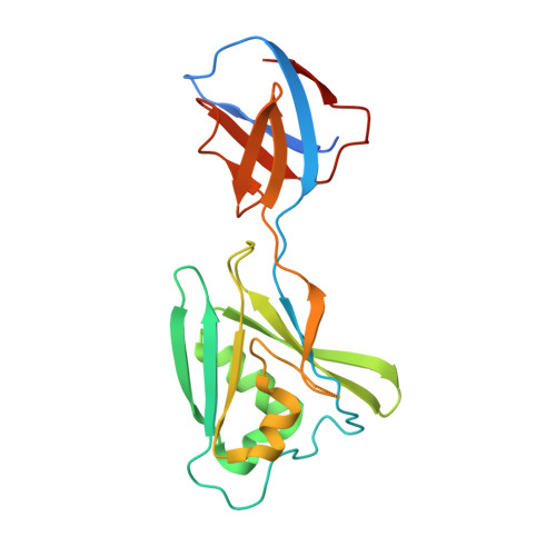
Tetrameric structure of the flagellar cap protein FliD from Serratia marcescens.

Cho, S.Y.Song, W.S.Hong, H.J.Lee, G.S.Kang, S.G.Ko, H.J.Kim, P.H.Yoon, S.I.

(2017) Biochem Biophys Res Commun 489: 63-69

  • DOI: https://doi.org/10.1016/j.bbrc.2017.05.093
  • Primary Citation Related Structures: 
    5XLJ, 5XLK

  • PubMed Abstract: 

    Bacterial motility is provided by the flagellum. FliD is located at the distal end of the flagellum and plays a key role in the insertion of each flagellin protein at the growing tip of the flagellar filament. Because FliD functions as an oligomer, the determination of the oligomeric state of FliD is critical to understanding the molecular mechanism of FliD-mediated flagellar growth. FliD has been shown to adopt a pentameric or a hexameric structure depending on the bacterial species. Here, we report another distinct oligomeric form of FliD based on structural and biochemical studies. The crystal structures of the D2 and D3 domains of Serratia marcescens FliD (smFliD) were determined in two crystal forms and together revealed that smFliD assembles into a tetrameric architecture that resembles a four-pointed star plate. smFliD tetramerization was also confirmed in solution by cross-linking experiments. Although smFliD oligomerizes in a head-to-tail orientation using a common primary binding interface between the D2 and D3' domains (the prime denotes the second subunit in the oligomer) similarly to other FliD orthologs, the smFliD tetramer diverges to present a unique secondary D2-D2' binding interface. Our structure-based comparative analysis of FliD suggests that bacteria have developed diverse species-specific oligomeric forms of FliD that range from tetramers to hexamers for flagellar growth.


  • Organizational Affiliation
    • Division of Biomedical Convergence, College of Biomedical Science, Kangwon National University, Chuncheon 24341, Republic of Korea.

Macromolecule Content 

  • Total Structure Weight: 66.05 kDa 
  • Atom Count: 4,007 
  • Modeled Residue Count: 563 
  • Deposited Residue Count: 624 
  • Unique protein chains: 1

Macromolecules

Find similar proteins by:|  3D Structure
Entity ID: 1
MoleculeChains  Sequence LengthOrganismDetailsImage
Flagellar hook-associated protein 2
A, B, C
208Serratia marcescensMutation(s): 0 
Gene Names: AR325_17470
UniProt
Find proteins for A0A0P0QFX8 (Serratia marcescens)
Explore A0A0P0QFX8 
Go to UniProtKB:  A0A0P0QFX8
Entity Groups
Sequence Clusters30% Identity50% Identity70% Identity90% Identity95% Identity100% Identity
UniProt GroupA0A0P0QFX8
Sequence Annotations
Expand
Reference Sequence

Small Molecules

Ligands 1 Unique
IDChains Name / Formula / InChI Key2D Diagram3D Interactions
ZN

Query on ZN


Download:Ideal Coordinates CCD File
D [auth A]
E [auth A]
F [auth A]
G [auth B]
H [auth B]
D [auth A],
E [auth A],
F [auth A],
G [auth B],
H [auth B],
I [auth B],
J [auth C],
K [auth C],
L [auth C]
ZINC ION
Zn
PTFCDOFLOPIGGS-UHFFFAOYSA-N

Experimental Data & Validation

Experimental Data

  • Method: X-RAY DIFFRACTION
  • Resolution: 3.05 Å
  • R-Value Free:  0.287 (Depositor), 0.286 (DCC) 
  • R-Value Work:  0.238 (Depositor), 0.238 (DCC) 
  • R-Value Observed: 0.241 (Depositor) 
Space Group: I 4 2 2
Unit Cell:
Length ( Å )Angle ( ˚ )
a = 121.362α = 90
b = 121.362β = 90
c = 249.266γ = 90
Software Package:
Software NamePurpose
REFMACrefinement
HKL-2000data reduction
HKL-2000data scaling
PHASERphasing

Structure Validation

View Full Validation Report


Entry History 

Deposition Data

Revision History  (Full details and data files)

  • Version 1.0: 2017-06-14
    Type: Initial release
  • Version 1.1: 2017-12-06
    Changes: Database references
  • Version 1.2: 2023-11-22
    Changes: Data collection, Database references, Derived calculations, Refinement description