5XFS

Crystal structure of PE8-PPE15 in complex with EspG5 from M. tuberculosis


Experimental Data Snapshot

  • Method: X-RAY DIFFRACTION
  • Resolution: 2.90 Å
  • R-Value Free: 0.262 
  • R-Value Work: 0.213 
  • R-Value Observed: 0.216 

wwPDB Validation   3D Report Full Report


This is version 1.2 of the entry. See complete history


Literature

Structural basis of the PE-PPE protein interaction in Mycobacterium tuberculosis.

Chen, X.Cheng, H.F.Zhou, J.Chan, C.Y.Lau, K.F.Tsui, S.K.Au, S.W.

(2017) J Biol Chem 292: 16880-16890

  • DOI: https://doi.org/10.1074/jbc.M117.802645
  • Primary Citation of Related Structures:  
    5XFS

  • PubMed Abstract: 

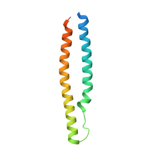
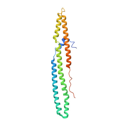
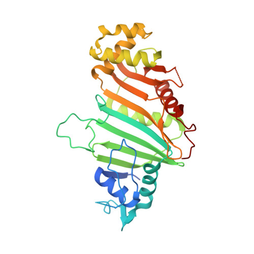
    Mycobacterium tuberculosis ( Mtb ), the causative agent of tuberculosis, has developed multiple strategies to adapt to the human host. The five type VII secretion systems, ESX-1-5, direct the export of many virulence-promoting protein effectors across the complex mycobacterial cell wall. One class of ESX substrates is the PE-PPE family of proteins, which is unique to mycobacteria and essential for infection, antigenic variation, and host-pathogen interactions. The genome of Mtb encodes 168 PE-PPE proteins. Many of them are thought to be secreted through ESX-5 secretion system and to function in pairs. However, understanding of the specific pairing of PE-PPE proteins and their structure-function relationship is limited by the challenging purification of many PE-PPE proteins, and our knowledge of the PE-PPE interactions therefore has been restricted to the PE25-PPE41 pair and its complex with the ESX-5 secretion system chaperone EspG5. Here, we report the crystal structure of a new PE-PPE pair, PE8-PPE15, in complex with EspG5. Our structure revealed that the EspG5-binding sites on PPE15 are relatively conserved among Mtb PPE proteins, suggesting that EspG5-PPE15 represents a more typical model for EspG5-PPE interactions than EspG5-PPE41. A structural comparison with the PE25-PPE41 complex disclosed conformational changes in the four-helix bundle structure and a unique binding mode in the PE8-PPE15 pair. Moreover, homology-modeling and mutagenesis studies further delineated the molecular determinants of the specific PE-PPE interactions. These findings help develop an atomic algorithm of ESX-5 substrate recognition and PE-PPE pairing.


  • Organizational Affiliation

    From the Centre for Protein Science and Crystallography, School of Life Sciences.


Macromolecules
Find similar proteins by:  (by identity cutoff)  |  3D Structure
Entity ID: 1
MoleculeChains Sequence LengthOrganismDetailsImage
PE family protein PE8104Mycobacterium tuberculosis H37RvMutation(s): 0 
Gene Names: PE8Rv1040c
UniProt
Find proteins for L7N667 (Mycobacterium tuberculosis (strain ATCC 25618 / H37Rv))
Explore L7N667 
Go to UniProtKB:  L7N667
Entity Groups  
Sequence Clusters30% Identity50% Identity70% Identity90% Identity95% Identity100% Identity
UniProt GroupL7N667
Sequence Annotations
Expand
  • Reference Sequence
Find similar proteins by:  (by identity cutoff)  |  3D Structure
Entity ID: 2
MoleculeChains Sequence LengthOrganismDetailsImage
PPE family protein PPE15202Mycobacterium tuberculosis H37RvMutation(s): 0 
Gene Names: PPE15mper1Rv1039c
UniProt
Find proteins for P9WI31 (Mycobacterium tuberculosis (strain ATCC 25618 / H37Rv))
Explore P9WI31 
Go to UniProtKB:  P9WI31
Entity Groups  
Sequence Clusters30% Identity50% Identity70% Identity90% Identity95% Identity100% Identity
UniProt GroupP9WI31
Sequence Annotations
Expand
  • Reference Sequence
Find similar proteins by:  (by identity cutoff)  |  3D Structure
Entity ID: 3
MoleculeChains Sequence LengthOrganismDetailsImage
ESX-5 secretion-associated protein EspG5308Mycobacterium tuberculosis H37RvMutation(s): 0 
Gene Names: espG5Rv1794LH57_09810
UniProt
Find proteins for O53943 (Mycobacterium tuberculosis (strain ATCC 25618 / H37Rv))
Explore O53943 
Go to UniProtKB:  O53943
Entity Groups  
Sequence Clusters30% Identity50% Identity70% Identity90% Identity95% Identity100% Identity
UniProt GroupO53943
Sequence Annotations
Expand
  • Reference Sequence
Experimental Data & Validation

Experimental Data

  • Method: X-RAY DIFFRACTION
  • Resolution: 2.90 Å
  • R-Value Free: 0.262 
  • R-Value Work: 0.213 
  • R-Value Observed: 0.216 
  • Space Group: P 21 21 21
Unit Cell:
Length ( Å )Angle ( ˚ )
a = 54.74α = 90
b = 69.96β = 90
c = 203.549γ = 90
Software Package:
Software NamePurpose
PHENIXrefinement
iMOSFLMdata processing

Structure Validation

View Full Validation Report


Entry History & Funding Information

Deposition Data


Funding OrganizationLocationGrant Number
Health and Medical Research FundHong Kong12110602

Revision History  (Full details and data files)

  • Version 1.0: 2017-08-30
    Type: Initial release
  • Version 1.1: 2017-12-06
    Changes: Database references
  • Version 1.2: 2023-11-22
    Changes: Data collection, Database references, Refinement description