5XFH

Crystal structure of Orysata lectin in complex with biantennary N-glycan


Experimental Data Snapshot

  • Method: X-RAY DIFFRACTION
  • Resolution: 1.90 Å
  • R-Value Free: 0.220 
  • R-Value Work: 0.189 
  • R-Value Observed: 0.191 

wwPDB Validation   3D Report Full Report


This is version 2.1 of the entry. See complete history


Literature

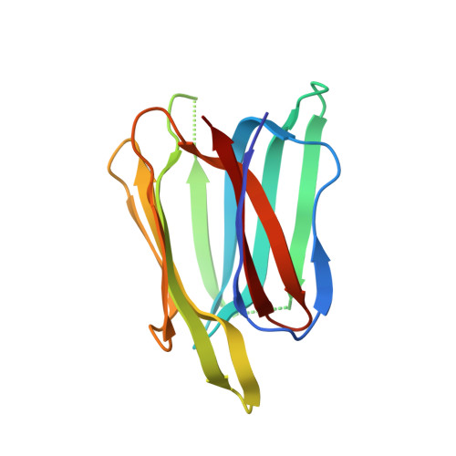
Distinct roles for each N-glycan branch interacting with mannose-binding type Jacalin-related lectins Orysata and Calsepa.

Nagae, M.Mishra, S.K.Hanashima, S.Tateno, H.Yamaguchi, Y.

(2017) Glycobiology 27: 1120-1133

  • DOI: https://doi.org/10.1093/glycob/cwx081
  • Primary Citation of Related Structures:  
    5XFH, 5XFI

  • PubMed Abstract: 

    Mannose-binding type Jacalin-related lectins (mJRLs) bind to branched N-glycans via conserved sugar-binding sites. Despite, significant 3D structural similarities, each mJRL is known to have a unique binding preference toward various N-glycans. However, the molecular basis of varying binding preference is substantially unknown. Here, we report a detailed comparison of N-glycan-binding preference for two mJRLs, Orysata and Calsepa using frontal affinity chromatography (FAC), X-ray and molecular modeling. The FAC analysis using a panel of N-glycans shows difference in N-glycan-binding preference between the lectins. Orysata shows broader specificity toward most high-mannose-type glycans as well as biantennary complex-type glycans bearing an extension on the Manα1-6 branch. Whereas, Calsepa shows narrow specificity to complex-type glycans with bisecting GlcNAc. The X-ray crystallographic structure reveals that two Orysata lectins bind to one biantennary N-glycan (2:1 binding) where one lectin binds to mannose of the α1-3 branch, while the other interacts with an N-acetylglucosamine of the α1-6 branch. In contrast, Calsepa shows 1:1 binding where α1-3 branch and core chitobiose region N-glycan interacts with lectin, while α1-6 branch is flipped-back to the chitobiose core. Molecular dynamics study of Orysata bound to N-glycans substantiate possibility of two-binding modes for each N-glycan. Binding free energies calculated separately for α1-3 and α1-6 branches of each N-glycan suggest both branches can bind to Orysata. Overall these results suggest that each branch of N-glycan has a distinct role in binding to mJRLs and the nonbinding branch can contribute significantly to the binding affinity and hence to the specificity.


  • Organizational Affiliation

    Structural Glycobiology Team, Systems Glycobiology Research Group, RIKEN Global Research Cluster, 2-1 Hirosawa, Wako, Saitama 351-0198, Japan.


Macromolecules
Find similar proteins by:  (by identity cutoff)  |  3D Structure
Entity ID: 1
MoleculeChains Sequence LengthOrganismDetailsImage
Salt stress-induced protein
A, B
145Oryza sativa Japonica GroupMutation(s): 0 
Gene Names: SALTMLOs01g0348900LOC_Os01g24710B1051E10.18P0440D10.39
UniProt
Find proteins for Q0JMY8 (Oryza sativa subsp. japonica)
Explore Q0JMY8 
Go to UniProtKB:  Q0JMY8
Entity Groups  
Sequence Clusters30% Identity50% Identity70% Identity90% Identity95% Identity100% Identity
UniProt GroupQ0JMY8
Sequence Annotations
Expand
  • Reference Sequence
Oligosaccharides

Help

Entity ID: 2
MoleculeChains Length2D Diagram Glycosylation3D Interactions
beta-D-galactopyranose-(1-4)-2-acetamido-2-deoxy-beta-D-glucopyranose-(1-2)-alpha-D-mannopyranose-(1-3)-[2-acetamido-2-deoxy-beta-D-glucopyranose-(1-2)-alpha-D-mannopyranose-(1-6)]beta-D-mannopyranose
C
6N/A
Glycosylation Resources
GlyTouCan:  G69611WV
GlyCosmos:  G69611WV
GlyGen:  G69611WV
Experimental Data & Validation

Experimental Data

  • Method: X-RAY DIFFRACTION
  • Resolution: 1.90 Å
  • R-Value Free: 0.220 
  • R-Value Work: 0.189 
  • R-Value Observed: 0.191 
  • Space Group: P 21 21 21
Unit Cell:
Length ( Å )Angle ( ˚ )
a = 45.783α = 90
b = 71.62β = 90
c = 97.201γ = 90
Software Package:
Software NamePurpose
PHENIXrefinement
HKL-2000data reduction
HKL-2000data scaling
MOLREPphasing

Structure Validation

View Full Validation Report


Entry History & Funding Information

Deposition Data


Funding OrganizationLocationGrant Number
MEXTJapan15K18496
MEXTJapan17K07303

Revision History  (Full details and data files)

  • Version 1.0: 2017-10-18
    Type: Initial release
  • Version 1.1: 2017-12-06
    Changes: Database references
  • Version 2.0: 2020-07-29
    Type: Remediation
    Reason: Carbohydrate remediation
    Changes: Atomic model, Data collection, Derived calculations, Structure summary
  • Version 2.1: 2023-11-22
    Changes: Data collection, Database references, Derived calculations, Refinement description, Structure summary