


Crystal structure of a hydrogen sulfide-producing enzyme (Fn1220) from Fusobacterium nucleatum in complex with L-lanthionine-PLP Schiff base
Kezuka, Y., Yoshida, Y., Nonaka, T.To be published.
Experimental Data Snapshot
Entity ID: 1 | |||||
|---|---|---|---|---|---|
| Molecule | Chains | Sequence Length | Organism | Details | Image |
| Cysteine synthase | 310 | Fusobacterium nucleatum subsp. nucleatum ATCC 25586 | Mutation(s): 0 Gene Names: FN1220 EC: 2.5.1.47 | 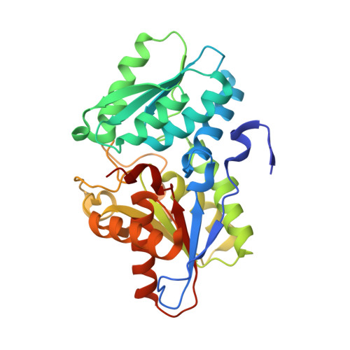 | |
UniProt | |||||
Entity Groups | |||||
| Sequence Clusters | 30% Identity50% Identity70% Identity90% Identity95% Identity100% Identity | ||||
| UniProt Group | Q8RE94 | ||||
Sequence AnnotationsExpand | |||||
Reference Sequence | |||||
| Ligands 7 Unique | |||||
|---|---|---|---|---|---|
| ID | Chains | Name / Formula / InChI Key | 2D Diagram | 3D Interactions | |
| PLP Download:Ideal Coordinates CCD File | D [auth A], P [auth B], R [auth B] | PYRIDOXAL-5'-PHOSPHATE C8 H10 N O6 P NGVDGCNFYWLIFO-UHFFFAOYSA-N |  | ||
| 85F Download:Ideal Coordinates CCD File | C [auth A], Q [auth B] | (2R)-2-azanyl-3-[(2R)-2-azanyl-3-oxidanyl-3-oxidanylidene-propyl]sulfanyl-propanoic acid C6 H12 N2 O4 S DWPCPZJAHOETAG-IMJSIDKUSA-N |  | ||
| PG5 Download:Ideal Coordinates CCD File | Z [auth B] | 1-METHOXY-2-[2-(2-METHOXY-ETHOXY]-ETHANE C8 H18 O4 YFNKIDBQEZZDLK-UHFFFAOYSA-N |  | ||
| PG0 Download:Ideal Coordinates CCD File | N [auth A] | 2-(2-METHOXYETHOXY)ETHANOL C5 H12 O3 SBASXUCJHJRPEV-UHFFFAOYSA-N |  | ||
| PEG Download:Ideal Coordinates CCD File | M [auth A], Y [auth B] | DI(HYDROXYETHYL)ETHER C4 H10 O3 MTHSVFCYNBDYFN-UHFFFAOYSA-N |  | ||
| ACT Download:Ideal Coordinates CCD File | E [auth A], F [auth A], S [auth B] | ACETATE ION C2 H3 O2 QTBSBXVTEAMEQO-UHFFFAOYSA-M |  | ||
| CA Download:Ideal Coordinates CCD File | G [auth A] H [auth A] I [auth A] J [auth A] K [auth A] | CALCIUM ION Ca BHPQYMZQTOCNFJ-UHFFFAOYSA-N |  | ||
| Length ( Å ) | Angle ( ˚ ) |
|---|---|
| a = 116.749 | α = 90 |
| b = 116.749 | β = 90 |
| c = 99.343 | γ = 90 |
| Software Name | Purpose |
|---|---|
| REFMAC | refinement |
| MOSFLM | data reduction |
| SCALA | data scaling |
| MOLREP | phasing |
| Funding Organization | Location | Grant Number |
|---|---|---|
| Japan Society for the Promotion of Science | Japan | JP21791810 |
| Japan Society for the Promotion of Science | Japan | JP23792130 |


