


Crystallographic study of homoserine dehydrogenase from Thermus thermophilus HB8
Akai, S., Ikushiro, H., Sawai, D., Hayashi, H., Kamiya, N., Miyahara, I.To be published.
Experimental Data Snapshot
wwPDB Validation 3D Report Full Report

Entity ID: 1 | |||||
|---|---|---|---|---|---|
| Molecule | Chains | Sequence Length | Organism | Details | Image |
| Homoserine dehydrogenase | 332 | Thermus thermophilus HB8 | Mutation(s): 0 Gene Names: TTHA0489 EC: 1.1.1.3 | 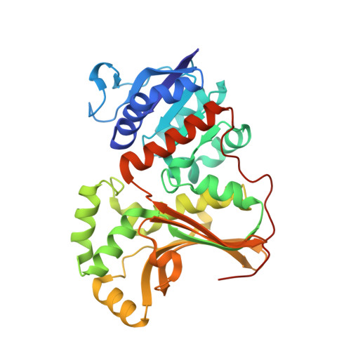 | |
UniProt | |||||
Entity Groups | |||||
| Sequence Clusters | 30% Identity50% Identity70% Identity90% Identity95% Identity100% Identity | ||||
| UniProt Group | Q5SL04 | ||||
Sequence AnnotationsExpand | |||||
Reference Sequence | |||||
| Ligands 3 Unique | |||||
|---|---|---|---|---|---|
| ID | Chains | Name / Formula / InChI Key | 2D Diagram | 3D Interactions | |
| HSE Download:Ideal Coordinates CCD File | C [auth A], F [auth B] | L-HOMOSERINE C4 H9 N O3 UKAUYVFTDYCKQA-VKHMYHEASA-N |  | ||
| FMT Download:Ideal Coordinates CCD File | E [auth A], H [auth B] | FORMIC ACID C H2 O2 BDAGIHXWWSANSR-UHFFFAOYSA-N |  | ||
| NA Download:Ideal Coordinates CCD File | D [auth A], G [auth B] | SODIUM ION Na FKNQFGJONOIPTF-UHFFFAOYSA-N |  | ||
| Length ( Å ) | Angle ( ˚ ) |
|---|---|
| a = 119.625 | α = 90 |
| b = 119.625 | β = 90 |
| c = 145.02 | γ = 120 |
| Software Name | Purpose |
|---|---|
| REFMAC | refinement |


