5X8G | pdb_00005x8g

Binary complex structure of a double mutant I454RA456K of o-Succinylbenzoate CoA Synthetase (MenE) from Bacillus Subtilis bound with its product analogue OSB-NCoA at 1.90 angstrom


Experimental Data Snapshot

  • Method: X-RAY DIFFRACTION
  • Resolution: 1.90 Å
  • R-Value Free: 
    0.206 (Depositor), 0.205 (DCC) 
  • R-Value Work: 
    0.164 (Depositor), 0.166 (DCC) 
  • R-Value Observed: 
    0.164 (Depositor) 

Starting Model: experimental
View more details

wwPDB Validation 3D Report Full Report

Validation slider image for 5X8G

Ligand Structure Quality Assessment 


This is version 1.3 of the entry. See complete history

Literature

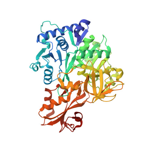
Crystal structure of the thioesterification conformation of Bacillus subtilis o-succinylbenzoyl-CoA synthetase reveals a distinct substrate-binding mode

Chen, Y.Li, T.L.Lin, X.Li, X.Li, X.D.Guo, Z.

(2017) J Biological Chem 292: 12296-12310

  • DOI: https://doi.org/10.1074/jbc.M117.790410
  • Primary Citation Related Structures: 
    5X8F, 5X8G

  • PubMed Abstract: 

    o -Succinylbenzoyl-CoA (OSB-CoA) synthetase (MenE) is an essential enzyme in bacterial vitamin K biosynthesis and an important target in the development of new antibiotics. It is a member of the adenylating enzymes (ANL) family, which reconfigure their active site in two different active conformations, one for the adenylation half-reaction and the other for a thioesterification half-reaction, in a domain-alternation catalytic mechanism. Although several aspects of the adenylating mechanism in MenE have recently been uncovered, its thioesterification conformation remains elusive. Here, using a catalytically competent Bacillus subtilis mutant protein complexed with an OSB-CoA analogue, we determined MenE high-resolution structures to 1.76 and 1.90 Å resolution in a thioester-forming conformation. By comparison with the adenylation conformation, we found that MenE's C-domain rotates around the Ser-384 hinge by 139.5° during domain-alternation catalysis. The structures also revealed a thioesterification active site specifically conserved among MenE orthologues and a substrate-binding mode distinct from those of many other acyl/aryl-CoA synthetases. Of note, using site-directed mutagenesis, we identified several residues that specifically contribute to the thioesterification half-reaction without affecting the adenylation half-reaction. Moreover, we observed a substantial movement of the activated succinyl group in the thioesterification half-reaction. These findings provide new insights into the domain-alternation catalysis of a bacterial enzyme essential for vitamin K biosynthesis and of its adenylating homologues in the ANL enzyme family.


  • Organizational Affiliation
    • Department of Chemistry, Hong Kong University of Science and Technology, Clear Water Bay, Kowloon, Hong Kong.

Macromolecule Content 

  • Total Structure Weight: 222.11 kDa 
  • Atom Count: 16,815 
  • Modeled Residue Count: 1,936 
  • Deposited Residue Count: 1,940 
  • Unique protein chains: 1

Macromolecules

Find similar proteins by:|  3D Structure
Entity ID: 1
MoleculeChains  Sequence LengthOrganismDetailsImage
2-succinylbenzoate--CoA ligaseA,
B,
C [auth D],
D [auth C]
485Bacillus subtilis subsp. subtilis str. 168Mutation(s): 2 
Gene Names: menEBSU30790
EC: 6.2.1.26
UniProt
Find proteins for P23971 (Bacillus subtilis (strain 168))
Explore P23971 
Go to UniProtKB:  P23971
Entity Groups
Sequence Clusters30% Identity50% Identity70% Identity90% Identity95% Identity100% Identity
UniProt GroupP23971
Sequence Annotations
Expand
Reference Sequence

Small Molecules

Ligands 5 Unique
IDChains Name / Formula / InChI Key2D Diagram3D Interactions
S0N

Query on S0N


Download:Ideal Coordinates CCD File
E [auth A],
JA [auth C],
O [auth B],
V [auth D]
o-succinylbenzoyl-N-coenzyme A
C32 H45 N8 O20 P3
QCTNXUGGWNSKFY-HSJNEKGZSA-N
PEG

Query on PEG


Download:Ideal Coordinates CCD File
AA [auth D]
BA [auth D]
HA [auth C]
IA [auth C]
L [auth A]
AA [auth D],
BA [auth D],
HA [auth C],
IA [auth C],
L [auth A],
M [auth A],
N [auth A]
DI(HYDROXYETHYL)ETHER
C4 H10 O3
MTHSVFCYNBDYFN-UHFFFAOYSA-N
CA

Query on CA


Download:Ideal Coordinates CCD File
CA [auth C]
DA [auth C]
EA [auth C]
FA [auth C]
I [auth A]
CA [auth C],
DA [auth C],
EA [auth C],
FA [auth C],
I [auth A],
J [auth A],
S [auth B],
Y [auth D]
CALCIUM ION
Ca
BHPQYMZQTOCNFJ-UHFFFAOYSA-N
MG

Query on MG


Download:Ideal Coordinates CCD File
F [auth A]
G [auth A]
H [auth A]
KA [auth C]
P [auth B]
F [auth A],
G [auth A],
H [auth A],
KA [auth C],
P [auth B],
Q [auth B],
R [auth B],
W [auth D],
X [auth D]
MAGNESIUM ION
Mg
JLVVSXFLKOJNIY-UHFFFAOYSA-N
NA

Query on NA


Download:Ideal Coordinates CCD File
GA [auth C],
K [auth A],
T [auth B],
U [auth B],
Z [auth D]
SODIUM ION
Na
FKNQFGJONOIPTF-UHFFFAOYSA-N

Experimental Data & Validation

Experimental Data

  • Method: X-RAY DIFFRACTION
  • Resolution: 1.90 Å
  • R-Value Free:  0.206 (Depositor), 0.205 (DCC) 
  • R-Value Work:  0.164 (Depositor), 0.166 (DCC) 
  • R-Value Observed: 0.164 (Depositor) 
Space Group: P 1
Unit Cell:
Length ( Å )Angle ( ˚ )
a = 71.95α = 80.14
b = 96.48β = 77.81
c = 97.66γ = 81.17
Software Package:
Software NamePurpose
PHENIXrefinement
Aimlessdata scaling
PDB_EXTRACTdata extraction
Aimlessdata reduction
PHENIXphasing

Structure Validation

View Full Validation Report


Ligand Structure Quality Assessment 


Entry History 

Deposition Data

Revision History  (Full details and data files)

  • Version 1.0: 2017-06-07
    Type: Initial release
  • Version 1.1: 2017-08-09
    Changes: Database references
  • Version 1.2: 2019-10-16
    Changes: Data collection, Derived calculations
  • Version 1.3: 2023-11-22
    Changes: Data collection, Database references, Derived calculations, Refinement description