5X6R | pdb_00005x6r

Crystal structure of Saccharomyces cerevisiae KMO in complex with Ro 61-8048


Experimental Data Snapshot

  • Method: X-RAY DIFFRACTION
  • Resolution: 1.91 Å
  • R-Value Free: 
    0.260 (Depositor), 0.260 (DCC) 
  • R-Value Work: 
    0.211 (Depositor), 0.213 (DCC) 
  • R-Value Observed: 
    0.212 (Depositor) 

Starting Model: experimental
View more details

wwPDB Validation 3D Report Full Report

Validation slider image for 5X6R

Ligand Structure Quality Assessment 


This is version 1.3 of the entry. See complete history

Literature

Structural Basis for Inhibitor-Induced Hydrogen Peroxide Production by Kynurenine 3-Monooxygenase

Kim, H.T.Na, B.K.Chung, J.Kim, S.Kwon, S.K.Cha, H.Son, J.Cho, J.M.Hwang, K.Y.

(2018) Cell Chem Biol 25: 426-438.e4

  • DOI: https://doi.org/10.1016/j.chembiol.2018.01.008
  • Primary Citation Related Structures: 
    5X68, 5X6P, 5X6Q, 5X6R

  • PubMed Abstract: 

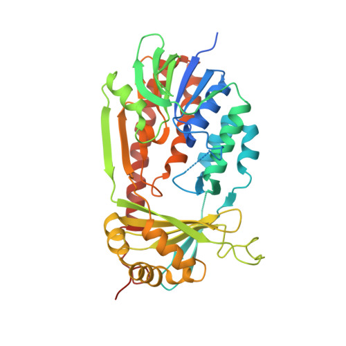
    Kynurenine 3-monooxygenase (KMO) inhibitors have been developed for the treatment of neurodegenerative disorders. The mechanisms of flavin reduction and hydrogen peroxide production by KMO inhibitors are unknown. Herein, we report the structure of human KMO and crystal structures of Saccharomyces cerevisiae (sc) and Pseudomonas fluorescens (pf) KMO with Ro 61-8048. Proton transfer in the hydrogen bond network triggers flavin reduction in p-hydroxybenzoate hydroxylase, but the mechanism triggering flavin reduction in KMO is different. Conformational changes via π-π interactions between the loop above the flavin and substrate or non-substrate effectors lead to disorder of the C-terminal α helix in scKMO and shifts of domain III in pfKMO, stimulating flavin reduction. Interestingly, Ro 61-8048 has two different binding modes. It acts as a competitive inhibitor in scKMO and as a non-substrate effector in pfKMO. These findings provide understanding of the catalytic cycle of KMO and insight for structure-based drug design of KMO inhibitors.


  • Organizational Affiliation
    • Crystalgenomics, Inc., 5F, Tower A, Korea Bio Park 700, Daewangpangyo-ro, Bundang-gu, Seongnam-si, Gyeonggi-do 13524, Korea; Division of Biotechnology, College of Life Sciences and Biotechnology, Korea University, 145 Anam-ro, Seongbuk-gu, Seoul 02841, Korea.

Macromolecule Content 

  • Total Structure Weight: 98.24 kDa 
  • Atom Count: 6,393 
  • Modeled Residue Count: 735 
  • Deposited Residue Count: 840 
  • Unique protein chains: 1

Macromolecules

Find similar proteins by:|  3D Structure
Entity ID: 1
MoleculeChains  Sequence LengthOrganismDetailsImage
Kynurenine 3-monooxygenase
A, B
420Saccharomyces cerevisiae S288CMutation(s): 0 
Gene Names: BNA4YBL098WYBL0828
EC: 1.14.13.9
UniProt
Find proteins for P38169 (Saccharomyces cerevisiae (strain ATCC 204508 / S288c))
Explore P38169 
Go to UniProtKB:  P38169
Entity Groups
Sequence Clusters30% Identity50% Identity70% Identity90% Identity95% Identity100% Identity
UniProt GroupP38169
Sequence Annotations
Expand
Reference Sequence

Experimental Data & Validation

Experimental Data

  • Method: X-RAY DIFFRACTION
  • Resolution: 1.91 Å
  • R-Value Free:  0.260 (Depositor), 0.260 (DCC) 
  • R-Value Work:  0.211 (Depositor), 0.213 (DCC) 
  • R-Value Observed: 0.212 (Depositor) 
Space Group: P 1 21 1
Unit Cell:
Length ( Å )Angle ( ˚ )
a = 58.878α = 90
b = 99.764β = 105.45
c = 84.693γ = 90
Software Package:
Software NamePurpose
PHENIXrefinement
HKL-2000data reduction
HKL-2000data scaling
MOLREPphasing

Structure Validation

View Full Validation Report


Ligand Structure Quality Assessment 


Entry History 

Deposition Data

Revision History  (Full details and data files)

  • Version 1.0: 2018-02-21
    Type: Initial release
  • Version 1.1: 2018-02-28
    Changes: Database references
  • Version 1.2: 2018-05-02
    Changes: Data collection, Database references
  • Version 1.3: 2023-11-22
    Changes: Data collection, Database references, Refinement description