


Crystal structure of a hydrolase encoded by lin2189 from Listeria innocua
Zhang, J., Zhou, J.To be published.
Experimental Data Snapshot
Starting Model: experimental
View more details
wwPDB Validation 3D Report Full Report

Entity ID: 1 | |||||
|---|---|---|---|---|---|
| Molecule | Chains | Sequence Length | Organism | Details | Image |
| Lin2189 protein | 216 | Listeria innocua Clip11262 | Mutation(s): 2 Gene Names: lin2189 | 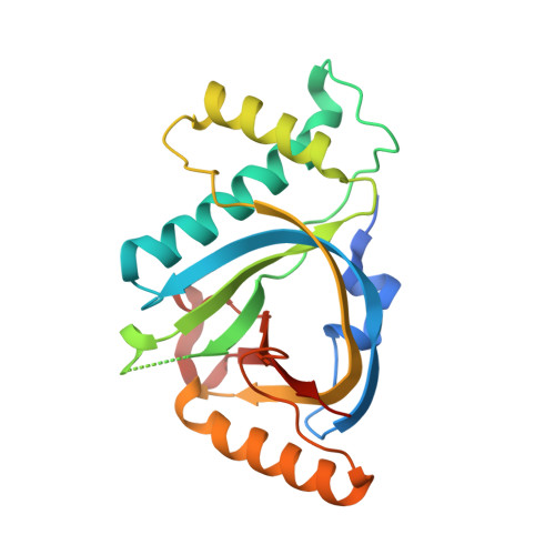 | |
UniProt | |||||
Entity Groups | |||||
| Sequence Clusters | 30% Identity50% Identity70% Identity90% Identity95% Identity100% Identity | ||||
| UniProt Group | Q929T5 | ||||
Sequence AnnotationsExpand | |||||
Reference Sequence | |||||
| Length ( Å ) | Angle ( ˚ ) |
|---|---|
| a = 120.011 | α = 90 |
| b = 48.759 | β = 125.51 |
| c = 80.301 | γ = 90 |
| Software Name | Purpose |
|---|---|
| PHENIX | refinement |
| HKL-3000 | data scaling |
| PHENIX | phasing |


