5X20 | pdb_00005x20

The ternary structure of D-mandelate dehydrogenase with NADH and anilino(oxo)acetate


Experimental Data Snapshot

  • Method: X-RAY DIFFRACTION
  • Resolution: 2.40 Å
  • R-Value Free: 
    0.218 (Depositor), 0.224 (DCC) 
  • R-Value Work: 
    0.184 (Depositor), 0.194 (DCC) 
  • R-Value Observed: 
    0.186 (Depositor) 

Starting Model: experimental
View more details

wwPDB Validation 3D Report Full Report

Validation slider image for 5X20

Ligand Structure Quality Assessment 


This is version 1.2 of the entry. See complete history

Literature

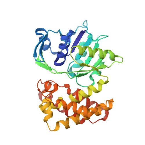
The ternary complex structure of d-mandelate dehydrogenase with NADH and anilino(oxo)acetate.

Furukawa, N.Miyanaga, A.Nakajima, M.Taguchi, H.

(2017) Biochem Biophys Res Commun 486: 665-670

  • DOI: https://doi.org/10.1016/j.bbrc.2017.03.088
  • Primary Citation Related Structures: 
    5X20

  • PubMed Abstract: 

    Enterococcus faecium NAD-dependent d-mandelate dehydrogenase (d-ManDH) belongs to a ketopantoate reductase (KPR)-related d-2-hydroxyacid dehydrogenase family, and exhibits broad substrate specificity toward bulky hydrophobic 2-ketoacids, preferring C3-branched substrates. The ternary complex structure of d-ManDH with NADH and anilino(oxo)acetate (AOA) revealed that the substrate binding induces a shear motion of the N-terminal domain along the C-terminal domain, following the hinge motion induced by the NADH binding, and allows the bound NADH molecule to form favorable interactions with a 2-ketoacid substrate. d-ManDH possesses a sufficiently wide pocket that accommodates the C3 branched side chains of substrates like KPR, but unlike the pocket of KPR, the pocket of d-ManDH comprises an entirely hydrophobic surface and an expanded space, in which the AOA benzene is accommodated. The expanded space mostly comprises a mobile loop structure, which likely modulates the shape and size of the space depending on the substrate.


  • Organizational Affiliation
    • Department of Applied Biological Science, Tokyo University of Science, 2641 Yamazaki, Noda, Chiba 278-8510, Japan.

Macromolecule Content 

  • Total Structure Weight: 213.76 kDa 
  • Atom Count: 13,694 
  • Modeled Residue Count: 1,671 
  • Deposited Residue Count: 1,872 
  • Unique protein chains: 1

Macromolecules

Find similar proteins by:|  3D Structure
Entity ID: 1
MoleculeChains  Sequence LengthOrganismDetailsImage
2-dehydropantoate 2-reductase
A, B, C, D, E
A, B, C, D, E, F
312Enterococcus faecium DOMutation(s): 0 
Gene Names: panEHMPREF0351_10295
EC: 1.1.1.169 (PDB Primary Data), 1.1.1.379 (UniProt), 1.1.1 (UniProt)
UniProt
Find proteins for Q3Y316 (Enterococcus faecium (strain ATCC BAA-472 / TX0016 / DO))
Explore Q3Y316 
Go to UniProtKB:  Q3Y316
Entity Groups
Sequence Clusters30% Identity50% Identity70% Identity90% Identity95% Identity100% Identity
UniProt GroupQ3Y316
Sequence Annotations
Expand
Reference Sequence

Small Molecules

Ligands 4 Unique
IDChains Name / Formula / InChI Key2D Diagram3D Interactions
NAD

Query on NAD


Download:Ideal Coordinates CCD File
AA [auth C],
G [auth A],
NA [auth D],
R [auth B],
SA [auth E]
NICOTINAMIDE-ADENINE-DINUCLEOTIDE
C21 H27 N7 O14 P2
BAWFJGJZGIEFAR-NNYOXOHSSA-N
AOT

Query on AOT


Download:Ideal Coordinates CCD File
BA [auth C],
H [auth A],
OA [auth D],
S [auth B],
TA [auth E]
2-oxidanylidene-2-phenylazanyl-ethanoic acid
C8 H7 N O3
PQJZHMCWDKOPQG-UHFFFAOYSA-N
SO4

Query on SO4


Download:Ideal Coordinates CCD File
GA [auth C]
HA [auth C]
IA [auth C]
JA [auth C]
KA [auth C]
GA [auth C],
HA [auth C],
IA [auth C],
JA [auth C],
KA [auth C],
LA [auth C],
MA [auth C],
N [auth A],
O [auth A],
P [auth A],
QA [auth D],
RA [auth D],
W [auth B],
X [auth B],
XA [auth E],
Y [auth B],
YA [auth E],
Z [auth B]
SULFATE ION
O4 S
QAOWNCQODCNURD-UHFFFAOYSA-L
GOL

Query on GOL


Download:Ideal Coordinates CCD File
CA [auth C]
DA [auth C]
EA [auth C]
FA [auth C]
I [auth A]
CA [auth C],
DA [auth C],
EA [auth C],
FA [auth C],
I [auth A],
J [auth A],
K [auth A],
L [auth A],
M [auth A],
PA [auth D],
Q [auth A],
T [auth B],
U [auth B],
UA [auth E],
V [auth B],
VA [auth E],
WA [auth E],
ZA [auth F]
GLYCEROL
C3 H8 O3
PEDCQBHIVMGVHV-UHFFFAOYSA-N

Experimental Data & Validation

Experimental Data

  • Method: X-RAY DIFFRACTION
  • Resolution: 2.40 Å
  • R-Value Free:  0.218 (Depositor), 0.224 (DCC) 
  • R-Value Work:  0.184 (Depositor), 0.194 (DCC) 
  • R-Value Observed: 0.186 (Depositor) 
Space Group: P 1 21 1
Unit Cell:
Length ( Å )Angle ( ˚ )
a = 96.3α = 90
b = 111.95β = 96.65
c = 108.94γ = 90
Software Package:
Software NamePurpose
REFMACrefinement
SCALAdata scaling
MOLREPphasing

Structure Validation

View Full Validation Report


Ligand Structure Quality Assessment 


Entry History 

Deposition Data

Revision History  (Full details and data files)

  • Version 1.0: 2017-04-05
    Type: Initial release
  • Version 1.1: 2017-12-06
    Changes: Data collection, Database references
  • Version 1.2: 2023-11-22
    Changes: Data collection, Database references, Refinement description