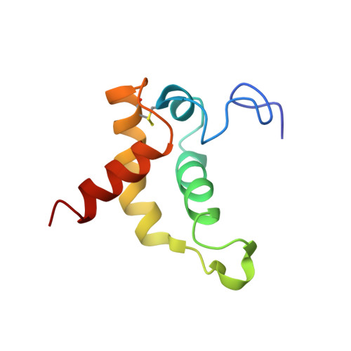
Characterizations of the Interactions between Escherichia coli Periplasmic Chaperone HdeA and Its Native Substrates during Acid Stress
Yu, X.C., Yang, C., Ding, J., Niu, X., Hu, Y., Jin, C.(2017) Biochemistry 56: 5748-5757
- PubMed: 29016106 Search on PubMed
- DOI: https://doi.org/10.1021/acs.biochem.7b00724
- Primary Citation Related Structures:
5WYO - PubMed Abstract:
The bacterial acid-resistant chaperone HdeA is a "conditionally disordered" protein that functions at low pH when it undergoes a transition from a well-folded dimer to an unfolded monomer. The dimer dissociation and unfolding processes result in exposure of hydrophobic surfaces that allows binding to a broad range of client proteins. To fully elucidate the chaperone mechanism of HdeA, it is crucial to understand how the activated HdeA interacts with its native substrates during acid stress. Herein, we present a nuclear magnetic resonance study of the pH-dependent HdeA-substrate interactions. Our results show that the activation of HdeA is not only induced by acidification but also regulated by the presence of unfolded substrates. The variable extent of unfolding of substrates differentially regulates the HdeA-substrate interaction, and the binding further affects the HdeA conformation. Finally, we show that HdeA binds its substrates heterogeneously, and the "amphiphilic" model for HdeA-substrate interaction is discussed.
- College of Chemistry and Molecular Engineering, ‡Beijing Nuclear Magnetic Resonance Center, §College of Life Sciences, and ∥Beijing National Laboratory for Molecular Sciences, Peking University , Beijing 100871, China.
Organizational Affiliation: