5WSU

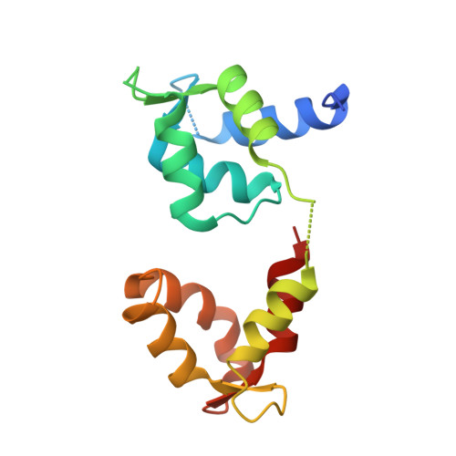
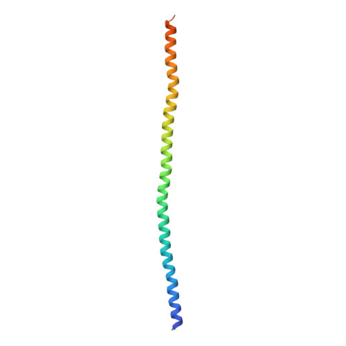
Crystal structure of Myosin VIIa IQ5-SAH in complex with apo-CaM


Experimental Data Snapshot

  • Method: X-RAY DIFFRACTION
  • Resolution: 3.00 Å
  • R-Value Free: 0.290 
  • R-Value Work: 0.224 
  • R-Value Observed: 0.231 

Starting Model: experimental
View more details

wwPDB Validation   3D Report Full Report


This is version 1.1 of the entry. See complete history


Literature

Ca(2+)-Induced Rigidity Change of the Myosin VIIa IQ Motif-Single alpha Helix Lever Arm Extension

Li, J.Chen, Y.Deng, Y.Unarta, I.C.Lu, Q.Huang, X.Zhang, M.

(2017) Structure 25: 579-591.e4

  • DOI: https://doi.org/10.1016/j.str.2017.02.002
  • Primary Citation of Related Structures:  
    5WST, 5WSU, 5WSV

  • PubMed Abstract: 

    Several unconventional myosins contain a highly charged single α helix (SAH) immediately following the calmodulin (CaM) binding IQ motifs, functioning to extend lever arms of these myosins. How such SAH is connected to the IQ motifs and whether the conformation of the IQ motifs-SAH segments are regulated by Ca 2+ fluctuations are not known. Here, we demonstrate by solving its crystal structure that the predicted SAH of myosin VIIa (Myo7a) forms a stable SAH. The structure of Myo7a IQ5-SAH segment in complex with apo-CaM reveals that the SAH sequence can extend the length of the Myo7a lever arm. Although Ca 2+ -CaM remains bound to IQ5-SAH, the Ca 2+ -induced CaM binding mode change softens the conformation of the IQ5-SAH junction, revealing a Ca 2+ -induced lever arm flexibility change for Myo7a. We further demonstrate that the last IQ motif of several other myosins also binds to both apo- and Ca 2+ -CaM, suggesting a common Ca 2+ -induced conformational regulation mechanism.


  • Organizational Affiliation

    Division of Life Science, State Key Laboratory of Molecular Neuroscience, Hong Kong University of Science and Technology, Clear Water Bay, Kowloon, Hong Kong, China. Electronic address: bcjli@connect.ust.hk.


Macromolecules
Find similar proteins by:  (by identity cutoff)  |  3D Structure
Entity ID: 1
MoleculeChains Sequence LengthOrganismDetailsImage
CalmodulinA,
C [auth B]
152Homo sapiensMutation(s): 0 
Gene Names: 
CALM1CALMCAMCAM1CALM2CAM2CAMBCALM3CALML2CAM3...
CALM1CALMCAMCAM1CALM2CAM2CAMBCALM3CALML2CAM3CAMCCAMIII

UniProt & NIH Common Fund Data Resources
Find proteins for P0DP23 (Homo sapiens)
Explore P0DP23 
Go to UniProtKB:  P0DP23
PHAROS:  P0DP23
GTEx:  ENSG00000198668 
Entity Groups  
Sequence Clusters30% Identity50% Identity70% Identity90% Identity95% Identity100% Identity
UniProt GroupP0DP23
Sequence Annotations
Expand
  • Reference Sequence
Find similar proteins by:  (by identity cutoff)  |  3D Structure
Entity ID: 2
MoleculeChains Sequence LengthOrganismDetailsImage
Unconventional myosin-VIIaB [auth C],
D
106Mus musculusMutation(s): 0 
Gene Names: Myo7aMyo7
UniProt & NIH Common Fund Data Resources
Find proteins for P97479 (Mus musculus)
Explore P97479 
Go to UniProtKB:  P97479
IMPC:  MGI:104510
Entity Groups  
Sequence Clusters30% Identity50% Identity70% Identity90% Identity95% Identity100% Identity
UniProt GroupP97479
Sequence Annotations
Expand
  • Reference Sequence
Experimental Data & Validation

Experimental Data

  • Method: X-RAY DIFFRACTION
  • Resolution: 3.00 Å
  • R-Value Free: 0.290 
  • R-Value Work: 0.224 
  • R-Value Observed: 0.231 
  • Space Group: P 41
Unit Cell:
Length ( Å )Angle ( ˚ )
a = 80.93α = 90
b = 80.93β = 90
c = 175.29γ = 90
Software Package:
Software NamePurpose
PHENIXrefinement
iMOSFLMdata reduction
SCALAdata scaling
MOLREPphasing

Structure Validation

View Full Validation Report


Entry History 

Deposition Data

Revision History  (Full details and data files)

  • Version 1.0: 2017-06-07
    Type: Initial release
  • Version 1.1: 2023-11-08
    Changes: Data collection, Database references, Refinement description