Discovery and Mechanistic Characterization of Selective Inhibitors of H2S-producing Enzyme: 3-Mercaptopyruvate Sulfurtransferase (3MST) Targeting Active-site Cysteine Persulfide
Hanaoka, K., Sasakura, K., Suwanai, Y., Toma-Fukai, S., Shimamoto, K., Takano, Y., Shibuya, N., Terai, T., Komatsu, T., Ueno, T., Ogasawara, Y., Tsuchiya, Y., Watanabe, Y., Kimura, H., Wang, C., Uchiyama, M., Kojima, H., Okabe, T., Urano, Y., Shimizu, T., Nagano, T.(2017) Sci Rep 7: 40227-40227
- PubMed: 28079151 Search on PubMedSearch on PubMed Central
- DOI: https://doi.org/10.1038/srep40227
- Primary Citation Related Structures:
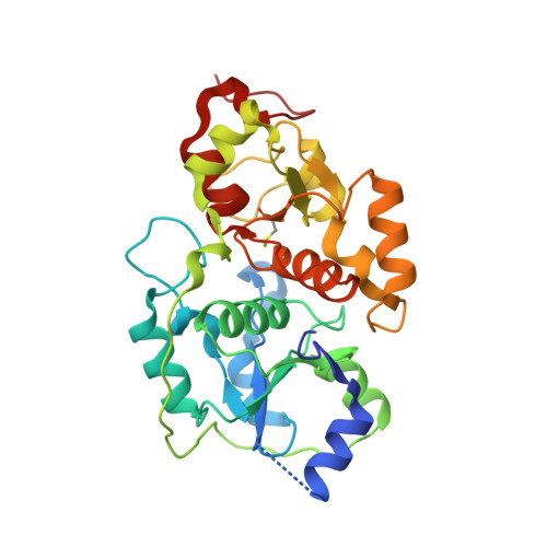
5WQJ, 5WQK - PubMed Abstract:
Very recent studies indicate that sulfur atoms with oxidation state 0 or -1, called sulfane sulfurs, are the actual mediators of some physiological processes previously considered to be regulated by hydrogen sulfide (H 2 S). 3-Mercaptopyruvate sulfurtransferase (3MST), one of three H 2 S-producing enzymes, was also recently shown to produce sulfane sulfur (H 2 S n ). Here, we report the discovery of several potent 3MST inhibitors by means of high-throughput screening (HTS) of a large chemical library (174,118 compounds) with our H 2 S-selective fluorescent probe, HSip-1. Most of the identified inhibitors had similar aromatic ring-carbonyl-S-pyrimidone structures. Among them, compound 3 showed very high selectivity for 3MST over other H 2 S/sulfane sulfur-producing enzymes and rhodanese. The X-ray crystal structures of 3MST complexes with two of the inhibitors revealed that their target is a persulfurated cysteine residue located in the active site of 3MST. Precise theoretical calculations indicated the presence of a strong long-range electrostatic interaction between the persulfur anion of the persulfurated cysteine residue and the positively charged carbonyl carbon of the pyrimidone moiety of the inhibitor. Our results also provide the experimental support for the idea that the 3MST-catalyzed reaction with 3-mercaptopyruvate proceeds via a ping-pong mechanism.
- Graduate School of Pharmaceutical Sciences, The University of Tokyo, 7-3-1 Hongo, Bunkyo-ku, Tokyo 113-0033, Japan.
Organizational Affiliation: