5WOP

High Resolution Structure of Mutant CA09-PB2cap


Experimental Data Snapshot

  • Method: X-RAY DIFFRACTION
  • Resolution: 1.52 Å
  • R-Value Free: 0.210 
  • R-Value Work: 0.148 
  • R-Value Observed: 0.151 

wwPDB Validation   3D Report Full Report


This is version 1.2 of the entry. See complete history


Literature

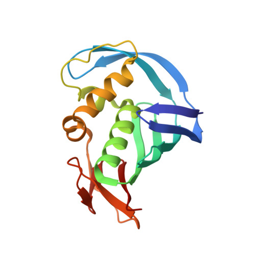
High-resolution structure of the Influenza A virus PB2cap binding domain illuminates the changes induced by ligand binding.

Constantinides, A.Gumpper, R.Severin, C.Luo, M.

(2018) Acta Crystallogr F Struct Biol Commun 74: 122-127

  • DOI: https://doi.org/10.1107/S2053230X18000894
  • Primary Citation of Related Structures:  
    5WOP

  • PubMed Abstract: 

    In the face of increasing drug resistance and the rapidly increasing necessity for practicality in clinical settings, drugs targeting different viral proteins are needed in order to control influenza A and B. A small molecule that tenaciously adheres to the PB2cap binding domain, part of the heterotrimeric RNA polymerase machinery of influenza A virus and influenza B virus, is a promising drug candidate. Understanding the anatomic behavior of PB2cap upon ligand binding will aid in the development of a more robust inhibitor. In this report, the anatomic behavior of the influenza A virus PB2cap domain is established by solving the crystal structure of native influenza A virus PB2cap at 1.52 Å resolution. By comparing it with the ligand-bound structure, the dissociation and rotation of the ligand-binding domain in PB2cap from the C-terminal domain is identified. This domain movement is present in many PB2cap structures, suggesting its functional relevance for polymerase activity.


  • Organizational Affiliation

    Department of Chemistry, Georgia State University, 50 Decatur Street, Atlanta, GA 30303, USA.


Macromolecules
Find similar proteins by:  (by identity cutoff)  |  3D Structure
Entity ID: 1
MoleculeChains Sequence LengthOrganismDetailsImage
Polymerase PB2
A, B
159Influenza A virusMutation(s): 0 
Gene Names: PB2
UniProt
Find proteins for F4MEU3 (Influenza A virus)
Explore F4MEU3 
Go to UniProtKB:  F4MEU3
Entity Groups  
Sequence Clusters30% Identity50% Identity70% Identity90% Identity95% Identity100% Identity
UniProt GroupF4MEU3
Sequence Annotations
Expand
  • Reference Sequence
Experimental Data & Validation

Experimental Data

  • Method: X-RAY DIFFRACTION
  • Resolution: 1.52 Å
  • R-Value Free: 0.210 
  • R-Value Work: 0.148 
  • R-Value Observed: 0.151 
  • Space Group: P 1 21 1
Unit Cell:
Length ( Å )Angle ( ˚ )
a = 36.863α = 90
b = 72.429β = 103.18
c = 61.316γ = 90
Software Package:
Software NamePurpose
REFMACrefinement
Aimlessdata scaling
PDB_EXTRACTdata extraction
MOLREPphasing
XDSdata reduction

Structure Validation

View Full Validation Report


Entry History 

Deposition Data

Revision History  (Full details and data files)

  • Version 1.0: 2017-12-20
    Type: Initial release
  • Version 1.1: 2018-03-14
    Changes: Database references
  • Version 1.2: 2023-10-04
    Changes: Data collection, Database references, Refinement description