5WJE

Crystal structure of Naa80 bound to a bisubstrate analogue


Experimental Data Snapshot

  • Method: X-RAY DIFFRACTION
  • Resolution: 1.76 Å
  • R-Value Free: 0.217 
  • R-Value Work: 0.177 
  • R-Value Observed: 0.181 

wwPDB Validation   3D Report Full Report


Ligand Structure Quality Assessment 


This is version 2.1 of the entry. See complete history


Literature

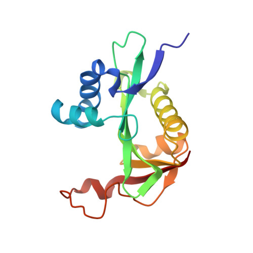
Structural determinants and cellular environment define processed actin as the sole substrate of the N-terminal acetyltransferase NAA80.

Goris, M.Magin, R.S.Foyn, H.Myklebust, L.M.Varland, S.Ree, R.Drazic, A.Bhambra, P.Stove, S.I.Baumann, M.Haug, B.E.Marmorstein, R.Arnesen, T.

(2018) Proc Natl Acad Sci U S A 115: 4405-4410

  • DOI: https://doi.org/10.1073/pnas.1719251115
  • Primary Citation of Related Structures:  
    5WJD, 5WJE

  • PubMed Abstract: 

    N-terminal (Nt) acetylation is a major protein modification catalyzed by N-terminal acetyltransferases (NATs). Methionine acidic N termini, including actin, are cotranslationally Nt acetylated by NatB in all eukaryotes, but animal actins containing acidic N termini, are additionally posttranslationally Nt acetylated by NAA80. Actin Nt acetylation was found to regulate cytoskeletal dynamics and motility, thus making NAA80 a potential target for cell migration regulation. In this work, we developed potent and selective bisubstrate inhibitors for NAA80 and determined the crystal structure of NAA80 in complex with such an inhibitor, revealing that NAA80 adopts a fold similar to other NAT enzymes but with a more open substrate binding region. Furthermore, in contrast to most other NATs, the substrate specificity of NAA80 is mainly derived through interactions between the enzyme and the acidic amino acids at positions 2 and 3 of the actin substrate and not residues 1 and 2. A yeast model revealed that ectopic expression of NAA80 in a strain lacking NatB activity partially restored Nt acetylation of NatB substrates, including yeast actin. Thus, NAA80 holds intrinsic capacity to posttranslationally Nt acetylate NatB-type substrates in vivo. In sum, the presence of a dominant cotranslational NatB in all eukaryotes, the specific posttranslational actin methionine removal in animals, and finally, the unique structural features of NAA80 leave only the processed actins as in vivo substrates of NAA80. Together, this study reveals the molecular and cellular basis of NAA80 Nt acetylation and provides a scaffold for development of inhibitors for the regulation of cytoskeletal properties.


  • Organizational Affiliation

    Department of Biological Sciences, University of Bergen, N-5020 Bergen, Norway.


Macromolecules
Find similar proteins by:  (by identity cutoff)  |  3D Structure
Entity ID: 1
MoleculeChains Sequence LengthOrganismDetailsImage
CG8481, isoform B159Drosophila melanogasterMutation(s): 0 
Gene Names: Dmel\CG8481CG8481-RBCG8481Dmel_CG8481
EC: 2.3.1
UniProt
Find proteins for Q59DX8 (Drosophila melanogaster)
Explore Q59DX8 
Go to UniProtKB:  Q59DX8
Entity Groups  
Sequence Clusters30% Identity50% Identity70% Identity90% Identity95% Identity100% Identity
UniProt GroupQ59DX8
Sequence Annotations
Expand
  • Reference Sequence

Find similar proteins by:  Sequence   |   3D Structure  

Entity ID: 2
MoleculeChains Sequence LengthOrganismDetailsImage
Actin N-terminus peptide5Drosophila melanogasterMutation(s): 0 
Sequence Annotations
Expand
  • Reference Sequence
Small Molecules
Ligands 1 Unique
IDChains Name / Formula / InChI Key2D Diagram3D Interactions
CMC
Query on CMC

Download Ideal Coordinates CCD File 
C [auth B]CARBOXYMETHYL COENZYME *A
C23 H38 N7 O18 P3 S
OBUOSIHPWVNVJN-GRFIIANRSA-N
Experimental Data & Validation

Experimental Data

  • Method: X-RAY DIFFRACTION
  • Resolution: 1.76 Å
  • R-Value Free: 0.217 
  • R-Value Work: 0.177 
  • R-Value Observed: 0.181 
  • Space Group: P 21 21 21
Unit Cell:
Length ( Å )Angle ( ˚ )
a = 37.136α = 90
b = 64.37β = 90
c = 68.589γ = 90
Software Package:
Software NamePurpose
PHENIXrefinement
HKL-2000data scaling
PDB_EXTRACTdata extraction
HKL-2000data reduction
PHENIXphasing

Structure Validation

View Full Validation Report


Ligand Structure Quality Assessment 


Entry History 

Deposition Data

Revision History  (Full details and data files)

  • Version 1.0: 2018-03-28
    Type: Initial release
  • Version 1.1: 2018-04-11
    Changes: Data collection, Database references
  • Version 1.2: 2018-05-02
    Changes: Data collection, Database references
  • Version 2.0: 2020-02-26
    Changes: Advisory, Atomic model, Data collection, Database references, Derived calculations, Polymer sequence, Source and taxonomy, Structure summary
  • Version 2.1: 2023-10-04
    Changes: Data collection, Database references, Refinement description