5WCZ | pdb_00005wcz

Crystal Structure of Wild-Type MalL from Bacillus subtilis with TS analogue 1-deoxynojirimycin


Experimental Data Snapshot

  • Method: X-RAY DIFFRACTION
  • Resolution: 1.58 Å
  • R-Value Free: 
    0.197 (Depositor), 0.172 (DCC) 
  • R-Value Work: 
    0.170 (Depositor), 0.172 (DCC) 
  • R-Value Observed: 
    0.171 (Depositor) 

Starting Model: experimental
View more details

wwPDB Validation 3D Report Full Report

Validation slider image for 5WCZ

Ligand Structure Quality Assessment 


This is version 1.2 of the entry. See complete history

Literature

Dynamical origins of heat capacity changes in enzyme-catalysed reactions.

van der Kamp, M.W.Prentice, E.J.Kraakman, K.L.Connolly, M.Mulholland, A.J.Arcus, V.L.

(2018) Nat Commun 9: 1177-1177

  • DOI: https://doi.org/10.1038/s41467-018-03597-y
  • Primary Citation Related Structures: 
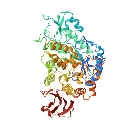
    5WCZ

  • PubMed Abstract: 

    Heat capacity changes are emerging as essential for explaining the temperature dependence of enzyme-catalysed reaction rates. This has important implications for enzyme kinetics, thermoadaptation and evolution, but the physical basis of these heat capacity changes is unknown. Here we show by a combination of experiment and simulation, for two quite distinct enzymes (dimeric ketosteroid isomerase and monomeric alpha-glucosidase), that the activation heat capacity change for the catalysed reaction can be predicted through atomistic molecular dynamics simulations. The simulations reveal subtle and surprising underlying dynamical changes: tightening of loops around the active site is observed, along with changes in energetic fluctuations across the whole enzyme including important contributions from oligomeric neighbours and domains distal to the active site. This has general implications for understanding enzyme catalysis and demonstrating a direct connection between functionally important microscopic dynamics and macroscopically measurable quantities.


  • Organizational Affiliation
    • School of Biochemistry, Biomedical Sciences Building, University of Bristol, University Walk, Bristol, BS8 1TD, UK. marc.vanderkamp@bristol.ac.uk.

Macromolecule Content 

  • Total Structure Weight: 139.07 kDa 
  • Atom Count: 10,395 
  • Modeled Residue Count: 1,110 
  • Deposited Residue Count: 1,172 
  • Unique protein chains: 1

Macromolecules

Find similar proteins by:|  3D Structure
Entity ID: 1
MoleculeChains  Sequence LengthOrganismDetailsImage
Oligo-1,6-glucosidase 1
A, B
586Bacillus subtilis subsp. subtilis str. 168Mutation(s): 0 
Gene Names: malLyvdLBSU34560
EC: 3.2.1.10
UniProt
Find proteins for O06994 (Bacillus subtilis (strain 168))
Explore O06994 
Go to UniProtKB:  O06994
Entity Groups
Sequence Clusters30% Identity50% Identity70% Identity90% Identity95% Identity100% Identity
UniProt GroupO06994
Sequence Annotations
Expand
Reference Sequence

Experimental Data & Validation

Experimental Data

  • Method: X-RAY DIFFRACTION
  • Resolution: 1.58 Å
  • R-Value Free:  0.197 (Depositor), 0.172 (DCC) 
  • R-Value Work:  0.170 (Depositor), 0.172 (DCC) 
  • R-Value Observed: 0.171 (Depositor) 
Space Group: P 1 21 1
Unit Cell:
Length ( Å )Angle ( ˚ )
a = 61.585α = 90
b = 98.835β = 103.08
c = 101.345γ = 90
Software Package:
Software NamePurpose
PHENIXrefinement
MOSFLMdata reduction
SCALAdata scaling
PDB_EXTRACTdata extraction
PHENIXphasing

Structure Validation

View Full Validation Report


Ligand Structure Quality Assessment 


Entry History 

Deposition Data

Revision History  (Full details and data files)

  • Version 1.0: 2018-03-28
    Type: Initial release
  • Version 1.1: 2018-04-04
    Changes: Data collection, Database references
  • Version 1.2: 2023-10-04
    Changes: Data collection, Database references, Refinement description