5WCG | pdb_00005wcg

SET and MYND Domain Containing protein 2


Experimental Data Snapshot

  • Method: X-RAY DIFFRACTION
  • Resolution: 2.02 Å
  • R-Value Free: 
    0.213 (Depositor), 0.223 (DCC) 
  • R-Value Work: 
    0.170 (Depositor), 0.180 (DCC) 
  • R-Value Observed: 
    0.171 (Depositor) 

Starting Model: experimental
View more details

wwPDB Validation 3D Report Full Report

Validation slider image for 5WCG

Ligand Structure Quality Assessment 


This is version 1.1 of the entry. See complete history

Literature

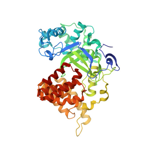
The crystal structure of SMYD2 in complex with compound MTF003

Zeng, H.Dong, A.Walker, J.R.Hutch, A.Seitova, A.Tatlock, J.Kumpf, R.Owen, A.Taylor, A.Casimiro-Garcia, A.Bountra, C.Arrowsmith, C.H.Edwards, A.M.Brown, P.J.Wu, H.

To be published.

Macromolecule Content 

  • Total Structure Weight: 50.74 kDa 
  • Atom Count: 3,696 
  • Modeled Residue Count: 430 
  • Deposited Residue Count: 433 
  • Unique protein chains: 1

Macromolecules

Find similar proteins by:|  3D Structure
Entity ID: 1
MoleculeChains  Sequence LengthOrganismDetailsImage
N-lysine methyltransferase SMYD2433Homo sapiensMutation(s): 0 
Gene Names: SMYD2KMT3C
EC: 2.1.1 (PDB Primary Data), 2.1.1.43 (PDB Primary Data), 2.1.1.354 (UniProt)
UniProt & NIH Common Fund Data Resources
Find proteins for Q9NRG4 (Homo sapiens)
Explore Q9NRG4 
Go to UniProtKB:  Q9NRG4
PHAROS:  Q9NRG4
GTEx:  ENSG00000143499 
Entity Groups
Sequence Clusters30% Identity50% Identity70% Identity90% Identity95% Identity100% Identity
UniProt GroupQ9NRG4
Sequence Annotations
Expand
Reference Sequence

Small Molecules

Ligands 5 Unique
IDChains Name / Formula / InChI Key2D Diagram3D Interactions
A4M
(Subject of Investigation/LOI)

Query on A4M


Download:Ideal Coordinates CCD File
E [auth A][3-(6-amino-2-methyl-9H-purin-9-yl)azetidin-1-yl]{5-[(4-cycloheptylpiperazin-1-yl)methyl]-1-methyl-1H-pyrazol-3-yl}methanone
C26 H38 N10 O
YBGLVGMKTGYRQH-UHFFFAOYSA-N
TRS

Query on TRS


Download:Ideal Coordinates CCD File
G [auth A]2-AMINO-2-HYDROXYMETHYL-PROPANE-1,3-DIOL
C4 H12 N O3
LENZDBCJOHFCAS-UHFFFAOYSA-O
DMS

Query on DMS


Download:Ideal Coordinates CCD File
F [auth A]DIMETHYL SULFOXIDE
C2 H6 O S
IAZDPXIOMUYVGZ-UHFFFAOYSA-N
ZN

Query on ZN


Download:Ideal Coordinates CCD File
B [auth A],
C [auth A],
D [auth A]
ZINC ION
Zn
PTFCDOFLOPIGGS-UHFFFAOYSA-N
UNX

Query on UNX


Download:Ideal Coordinates CCD File
H [auth A],
I [auth A],
J [auth A],
K [auth A],
L [auth A]
UNKNOWN ATOM OR ION
X
Binding Affinity Annotations 
IDSourceBinding Affinity
A4M BindingDB:  5WCG IC50: min: 40, max: 7000 (nM) from 2 assay(s)

Experimental Data & Validation

Experimental Data

  • Method: X-RAY DIFFRACTION
  • Resolution: 2.02 Å
  • R-Value Free:  0.213 (Depositor), 0.223 (DCC) 
  • R-Value Work:  0.170 (Depositor), 0.180 (DCC) 
  • R-Value Observed: 0.171 (Depositor) 
Space Group: C 1 2 1
Unit Cell:
Length ( Å )Angle ( ˚ )
a = 92.343α = 90
b = 98.312β = 101.95
c = 58.833γ = 90
Software Package:
Software NamePurpose
SCALEPACKdata scaling
PHASERphasing
REFMACrefinement
PDB_EXTRACTdata extraction
HKL-3000data reduction
HKL-3000phasing

Structure Validation

View Full Validation Report


Ligand Structure Quality Assessment 


Entry History 

Revision History  (Full details and data files)

  • Version 1.0: 2017-08-02
    Type: Initial release
  • Version 1.1: 2023-10-04
    Changes: Data collection, Database references, Derived calculations, Refinement description