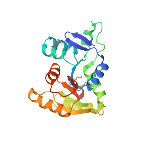
5WB4

Crystal structure of the TarA wall teichoic acid glycosyltransferase


Experimental Data Snapshot

  • Method: X-RAY DIFFRACTION
  • Resolution: 2.00 Å
  • R-Value Free: 0.246 
  • R-Value Work: 0.219 
  • R-Value Observed: 0.221 

wwPDB Validation   3D Report Full Report


This is version 1.2 of the entry. See complete history


Literature

Structure and mechanism of TagA, a novel membrane-associated glycosyltransferase that produces wall teichoic acids in pathogenic bacteria.

Kattke, M.D.Gosschalk, J.E.Martinez, O.E.Kumar, G.Gale, R.T.Cascio, D.Sawaya, M.R.Philips, M.Brown, E.D.Clubb, R.T.

(2019) PLoS Pathog 15: e1007723-e1007723

  • DOI: https://doi.org/10.1371/journal.ppat.1007723
  • Primary Citation of Related Structures:  
    5WB4, 5WFG

  • PubMed Abstract: 

    Staphylococcus aureus and other bacterial pathogens affix wall teichoic acids (WTAs) to their surface. These highly abundant anionic glycopolymers have critical functions in bacterial physiology and their susceptibility to β-lactam antibiotics. The membrane-associated TagA glycosyltransferase (GT) catalyzes the first-committed step in WTA biosynthesis and is a founding member of the WecB/TagA/CpsF GT family, more than 6,000 enzymes that synthesize a range of extracellular polysaccharides through a poorly understood mechanism. Crystal structures of TagA from T. italicus in its apo- and UDP-bound states reveal a novel GT fold, and coupled with biochemical and cellular data define the mechanism of catalysis. We propose that enzyme activity is regulated by interactions with the bilayer, which trigger a structural change that facilitates proper active site formation and recognition of the enzyme's lipid-linked substrate. These findings inform upon the molecular basis of WecB/TagA/CpsF activity and could guide the development of new anti-microbial drugs.


  • Organizational Affiliation

    Department of Chemistry and Biochemistry, University of California, Los Angeles, Los Angeles, United States of America.


Macromolecules
Find similar proteins by:  (by identity cutoff)  |  3D Structure
Entity ID: 1
MoleculeChains Sequence LengthOrganismDetailsImage
N-acetylglucosaminyldiphosphoundecaprenol N-acetyl-beta-D-mannosaminyltransferase
A, B, C, D, E
A, B, C, D, E, F, G, H
195Thermoanaerobacter italicus Ab9Mutation(s): 1 
Gene Names: Thit_1850
EC: 2.4.1.187
UniProt
Find proteins for D3T4E0 (Thermoanaerobacter italicus (strain DSM 9252 / Ab9))
Explore D3T4E0 
Go to UniProtKB:  D3T4E0
Entity Groups  
Sequence Clusters30% Identity50% Identity70% Identity90% Identity95% Identity100% Identity
UniProt GroupD3T4E0
Sequence Annotations
Expand
  • Reference Sequence
Small Molecules
Ligands 1 Unique
IDChains Name / Formula / InChI Key2D Diagram3D Interactions
SO4
Query on SO4

Download Ideal Coordinates CCD File 
I [auth A]
J [auth B]
K [auth B]
L [auth B]
M [auth C]
I [auth A],
J [auth B],
K [auth B],
L [auth B],
M [auth C],
N [auth E],
O [auth F],
P [auth G],
Q [auth H]
SULFATE ION
O4 S
QAOWNCQODCNURD-UHFFFAOYSA-L
Modified Residues  1 Unique
IDChains TypeFormula2D DiagramParent
MSE
Query on MSE
A, B, C, D, E
A, B, C, D, E, F, G, H
L-PEPTIDE LINKINGC5 H11 N O2 SeMET
Experimental Data & Validation

Experimental Data

  • Method: X-RAY DIFFRACTION
  • Resolution: 2.00 Å
  • R-Value Free: 0.246 
  • R-Value Work: 0.219 
  • R-Value Observed: 0.221 
  • Space Group: P 1 21 1
Unit Cell:
Length ( Å )Angle ( ˚ )
a = 77.03α = 90
b = 107.79β = 98.29
c = 89.44γ = 90
Software Package:
Software NamePurpose
XSCALEdata scaling
BUSTERrefinement
PDB_EXTRACTdata extraction
XSCALEdata reduction
SHELXDEphasing

Structure Validation

View Full Validation Report


Entry History 

Deposition Data

Revision History  (Full details and data files)

  • Version 1.0: 2019-01-16
    Type: Initial release
  • Version 1.1: 2019-07-31
    Changes: Data collection, Database references
  • Version 1.2: 2024-10-16
    Changes: Data collection, Database references, Structure summary