Structure-Based Analysis of Boronic Acids as Inhibitors of Acinetobacter-Derived Cephalosporinase-7, a Unique Class C beta-Lactamase.
Bouza, A.A., Swanson, H.C., Smolen, K.A., VanDine, A.L., Taracila, M.A., Romagnoli, C., Caselli, E., Prati, F., Bonomo, R.A., Powers, R.A., Wallar, B.J.(2018) ACS Infect Dis 4: 325-336
- PubMed: 29144724 Search on PubMedSearch on PubMed Central
- DOI: https://doi.org/10.1021/acsinfecdis.7b00152
- Primary Citation Related Structures:
5WAC, 5WAD, 5WAE, 5WAF, 5WAG - PubMed Abstract:
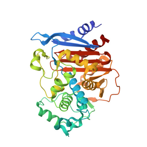
Acinetobacter baumannii is a multidrug resistant pathogen that infects more than 12 000 patients each year in the US. Much of the resistance to β-lactam antibiotics in Acinetobacter spp. is mediated by class C β-lactamases known as Acinetobacter-derived cephalosporinases (ADCs). ADCs are unaffected by clinically used β-lactam-based β-lactamase inhibitors. In this study, five boronic acid transition state analog inhibitors (BATSIs) were evaluated for inhibition of the class C cephalosporinase ADC-7. Our goal was to explore the properties of BATSIs designed to probe the R1 binding site. K i values ranged from low micromolar to subnanomolar, and circular dichroism (CD) demonstrated that each inhibitor stabilizes the β-lactamase-inhibitor complexes. Additionally, X-ray crystal structures of ADC-7 in complex with five inhibitors were determined (resolutions from 1.80 to 2.09 Å). In the ADC-7/CR192 complex, the BATSI with the lowest K i (0.45 nM) and greatest Δ T m (+9 °C), a trifluoromethyl substituent, interacts with Arg340. Arg340 is unique to ADCs and may play an important role in the inhibition of ADC-7. The ADC-7/BATSI complexes determined in this study shed light into the unique recognition sites in ADC enzymes and also offer insight into further structure-based optimization of these inhibitors.
- Department of Chemistry , Grand Valley State University , 1 Campus Drive , Allendale , Michigan 49401 , United States.
Organizational Affiliation: