5W6J | pdb_00005w6j

Agrobacterium tumefaciens ADP-glucose pyrophosphorylase


Experimental Data Snapshot

  • Method: X-RAY DIFFRACTION
  • Resolution: 1.78 Å
  • R-Value Free: 
    0.229 (Depositor) 
  • R-Value Work: 
    0.200 (Depositor), 0.174 (DCC) 
  • R-Value Observed: 
    0.202 (Depositor) 

wwPDB Validation 3D Report Full Report

Validation slider image for 5W6J

This is version 1.4 of the entry. See complete history

Literature

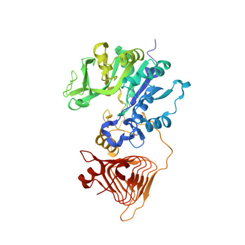
Structural analysis reveals a pyruvate-binding activator site in theAgrobacterium tumefaciensADP-glucose pyrophosphorylase.

Hill, B.L.Mascarenhas, R.Patel, H.P.Asencion Diez, M.D.Wu, R.Iglesias, A.A.Liu, D.Ballicora, M.A.

(2019) J Biological Chem 294: 1338-1348

  • DOI: https://doi.org/10.1074/jbc.RA118.004246
  • Primary Citation Related Structures: 
    5W5R, 5W5T, 5W6J

  • PubMed Abstract: 

    The pathways for biosynthesis of glycogen in bacteria and starch in plants are evolutionarily and biochemically related. They are regulated primarily by ADP-glucose pyrophosphorylase, which evolved to satisfy metabolic requirements of a particular organism. Despite the importance of these two pathways, little is known about the mechanism that controls pyrophosphorylase activity or the location of its allosteric sites. Here, we report pyruvate-bound crystal structures of ADP-glucose pyrophosphorylase from the bacterium Agrobacterium tumefaciens , identifying a previously elusive activator site for the enzyme. We found that the tetrameric enzyme binds two molecules of pyruvate in a planar conformation. Each binding site is located in a crevice between the C-terminal domains of two subunits where they stack via a distinct β-helix region. Pyruvate interacts with the side chain of Lys-43 and with the peptide backbone of Ser-328 and Gly-329 from both subunits. These structural insights led to the design of two variants with altered regulatory properties. In one variant (K43A), the allosteric effect was absent, whereas in the other (G329D), the introduced Asp mimicked the presence of pyruvate. The latter generated an enzyme that was preactivated and insensitive to further activation by pyruvate. Our study furnishes a deeper understanding of how glycogen biosynthesis is regulated in bacteria and the mechanism by which transgenic plants increased their starch production. These insights will facilitate rational approaches to enzyme engineering for starch production in crops of agricultural interest and will promote further study of allosteric signal transmission and molecular evolution in this important enzyme family.


  • Organizational Affiliation
    • Department of Chemistry and Biochemistry, Loyola University Chicago, Chicago, Illinois 60660.

Macromolecule Content 

  • Total Structure Weight: 946.06 kDa 
  • Atom Count: 75,925 
  • Modeled Residue Count: 8,228 
  • Deposited Residue Count: 8,400 
  • Unique protein chains: 1

Macromolecules

Find similar proteins by:|  3D Structure
Entity ID: 1
MoleculeChains  Sequence LengthOrganismDetailsImage
Glucose-1-phosphate adenylyltransferase420Agrobacterium tumefaciensMutation(s): 0 
Gene Names: glgC
EC: 2.7.7.27
UniProt
Find proteins for P39669 (Agrobacterium tumefaciens)
Explore P39669 
Go to UniProtKB:  P39669
Entity Groups
Sequence Clusters30% Identity50% Identity70% Identity90% Identity95% Identity100% Identity
UniProt GroupP39669
Sequence Annotations
Expand
Reference Sequence

Small Molecules

Ligands 1 Unique
IDChains Name / Formula / InChI Key2D Diagram3D Interactions
SO4

Query on SO4


Download:Ideal Coordinates CCD File
AA [auth D]
AB [auth F]
BA [auth D]
BB [auth G]
CA [auth E]
AA [auth D],
AB [auth F],
BA [auth D],
BB [auth G],
CA [auth E],
CB [auth G],
DA [auth E],
DB [auth M],
EA [auth E],
EB [auth M],
FA [auth H],
FB [auth T],
GA [auth H],
GB [auth T],
HA [auth I],
HB [auth T],
IA [auth I],
IB [auth U],
JA [auth J],
JB [auth U],
KA [auth J],
LA [auth K],
MA [auth K],
NA [auth L],
OA [auth L],
PA [auth N],
QA [auth N],
RA [auth O],
SA [auth O],
TA [auth P],
U [auth A],
UA [auth P],
V [auth A],
VA [auth Q],
W [auth B],
WA [auth Q],
X [auth B],
XA [auth R],
Y [auth C],
YA [auth R],
Z [auth C],
ZA [auth F]
SULFATE ION
O4 S
QAOWNCQODCNURD-UHFFFAOYSA-L

Experimental Data & Validation

Experimental Data

  • Method: X-RAY DIFFRACTION
  • Resolution: 1.78 Å
  • R-Value Free:  0.229 (Depositor) 
  • R-Value Work:  0.200 (Depositor), 0.174 (DCC) 
  • R-Value Observed: 0.202 (Depositor) 
Space Group: P 1
Unit Cell:
Length ( Å )Angle ( ˚ )
a = 93.439α = 72.04
b = 140.685β = 78.19
c = 228.49γ = 89.98
Software Package:
Software NamePurpose
PHENIXrefinement
HKL-3000data scaling
PDB_EXTRACTdata extraction
HKL-3000data reduction
PHASERphasing

Structure Validation

View Full Validation Report


Entry History 

& Funding Information

Deposition Data


Funding OrganizationLocationGrant Number
National Science Foundation (NSF, United States)United StatesMCB1616851

Revision History  (Full details and data files)

  • Version 1.0: 2018-11-14
    Type: Initial release
  • Version 1.1: 2018-11-21
    Changes: Data collection, Database references
  • Version 1.2: 2019-02-20
    Changes: Author supporting evidence, Data collection
  • Version 1.3: 2019-11-27
    Changes: Author supporting evidence, Database references
  • Version 1.4: 2024-03-13
    Changes: Advisory, Data collection, Database references