5W4S | pdb_00005w4s

Solution structure of C2 domain from protein kinase C alpha in ternary complex with calcium and V5-pHM peptide

  • Classification: TRANSFERASE
  • Organism(s): Rattus norvegicus
  • Expression System: Escherichia coli
  • Mutation(s): No 

  • Deposited: 2017-06-12 Released: 2018-04-25 
  • Deposition Author(s): Yang, Y., Igumenova, T.I.
  • Funding Organization(s): Welch Foundation, National Institutes of Health/National Institute of General Medical Sciences (NIH/NIGMS), National Science Foundation (NSF, United States)

Experimental Data Snapshot

  • Method: SOLUTION NMR
  • Conformers Calculated: 400 
  • Conformers Submitted: 20 
  • Selection Criteria: structures with the lowest energy 

wwPDB Validation 3D Report Full Report

Validation slider image for 5W4S

This is version 1.3 of the entry. See complete history

Literature

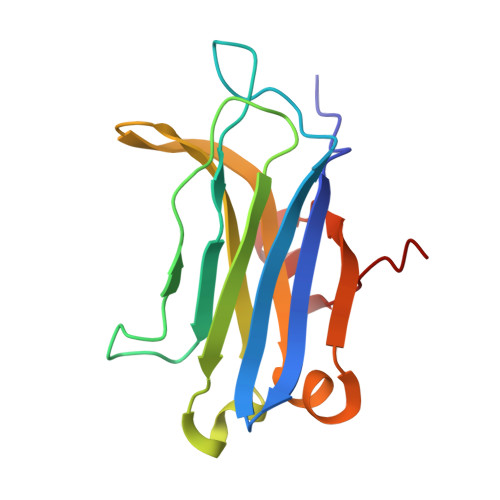
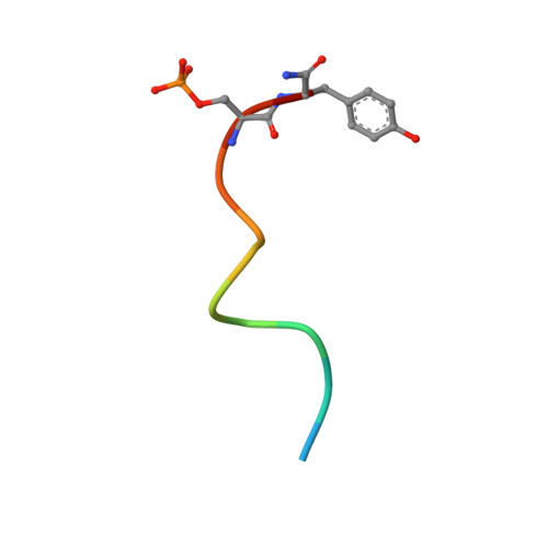
Structural Basis of Protein Kinase C alpha Regulation by the C-Terminal Tail.

Yang, Y.Shu, C.Li, P.Igumenova, T.I.

(2018) Biophys J 114: 1590-1603

  • DOI: https://doi.org/10.1016/j.bpj.2017.12.030
  • Primary Citation Related Structures: 
    5W4S

  • PubMed Abstract: 

    Protein kinase C (PKC) isoenzymes are multi-modular proteins activated at the membrane surface to regulate signal transduction processes. When activated by second messengers, PKC undergoes a drastic conformational and spatial transition from the inactive cytosolic state to the activated membrane-bound state. The complete structure of either state of PKC remains elusive. We demonstrate, using NMR spectroscopy, that the isolated Ca 2+ -sensing membrane-binding C2 domain of the conventional PKCα interacts with a conserved hydrophobic motif of the kinase C-terminal region, and we report a structural model of the complex. Our data suggest that the C-terminal region plays a dual role in regulating the PKC activity: activating, through sensitization of PKC to intracellular Ca 2+ oscillations; and auto-inhibitory, through its interaction with a conserved positively charged region of the C2 domain.


  • Organizational Affiliation
    • Department of Biochemistry and Biophysics, Texas A&M University, College Station, Texas.

Macromolecule Content 

  • Total Structure Weight: 17.62 kDa 
  • Atom Count: 1,237 
  • Modeled Residue Count: 151 
  • Deposited Residue Count: 151 
  • Unique protein chains: 2

Macromolecules

Find similar proteins by:|  3D Structure
Entity ID: 1
MoleculeChains  Sequence LengthOrganismDetailsImage
Protein kinase C alpha type139Rattus norvegicusMutation(s): 0 
Gene Names: PrkcaPkca
EC: 2.7.11.13
UniProt
Find proteins for P05696 (Rattus norvegicus)
Explore P05696 
Go to UniProtKB:  P05696
Entity Groups
Sequence Clusters30% Identity50% Identity70% Identity90% Identity95% Identity100% Identity
UniProt GroupP05696
Sequence Annotations
Expand
Reference Sequence
Find similar proteins by:  Sequence   |   3D Structure  
Entity ID: 2
MoleculeChains  Sequence LengthOrganismDetailsImage
V5-pHM peptide12Rattus norvegicusMutation(s): 0 
EC: 2.7.11.13
UniProt
Find proteins for P05696 (Rattus norvegicus)
Explore P05696 
Go to UniProtKB:  P05696
Entity Groups
Sequence Clusters30% Identity50% Identity70% Identity90% Identity95% Identity100% Identity
UniProt GroupP05696
Sequence Annotations
Expand
Reference Sequence

Small Molecules

Modified Residues  1 Unique
IDChains TypeFormula2D DiagramParent
SEP
Query on SEP
B
L-PEPTIDE LINKINGC3 H8 N O6 PSER

Experimental Data & Validation

Experimental Data

  • Method: SOLUTION NMR
  • Conformers Calculated: 400 
  • Conformers Submitted: 20 
  • Selection Criteria: structures with the lowest energy 

Structure Validation

View Full Validation Report


Entry History 

& Funding Information

Deposition Data


Funding OrganizationLocationGrant Number
Welch FoundationUnited StatesA-1784
National Institutes of Health/National Institute of General Medical Sciences (NIH/NIGMS)United StatesGM108998
National Science Foundation (NSF, United States)United StatesCHE-1151435

Revision History  (Full details and data files)

  • Version 1.0: 2018-04-25
    Type: Initial release
  • Version 1.1: 2019-11-27
    Changes: Author supporting evidence, Data collection
  • Version 1.2: 2023-06-14
    Changes: Database references, Derived calculations, Other
  • Version 1.3: 2024-10-30
    Changes: Data collection, Database references, Structure summary