5W4M

Crystal structure of Streptococcus dysgalactiae SHP pheromone receptor Rgg2(C45S)


Experimental Data Snapshot

  • Method: X-RAY DIFFRACTION
  • Resolution: 2.39 Å
  • R-Value Free: 0.224 
  • R-Value Work: 0.193 
  • R-Value Observed: 0.195 

wwPDB Validation   3D Report Full Report


This is version 1.5 of the entry. See complete history


Literature

Activating mutations in quorum-sensing regulator Rgg2 and its conformational flexibility in the absence of an intermolecular disulfide bond.

Wilkening, R.V.Capodagli, G.C.Khataokar, A.Tylor, K.M.Neiditch, M.B.Federle, M.J.

(2017) J Biol Chem 292: 20544-20557

  • DOI: https://doi.org/10.1074/jbc.M117.801670
  • Primary Citation of Related Structures:  
    5W4M, 5W4N

  • PubMed Abstract: 

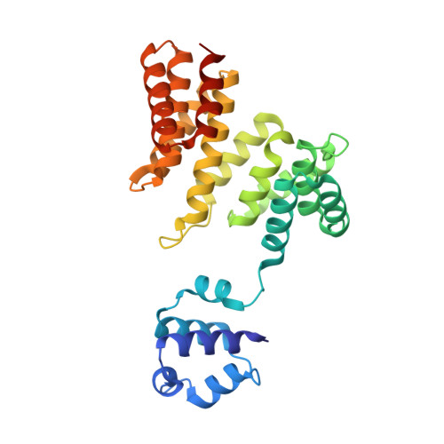
    Rap/Rgg/NprR/PlcR/PrgX (RRNPP) quorum-sensing systems use extracellular peptide pheromones that are detected by cytoplasmic receptors to regulate gene expression in firmicute bacteria. Rgg-type receptors are allosterically regulated through direct pheromone binding to control transcriptional activity; however, the receptor activation mechanism remains poorly understood. Previous work has identified a disulfide bond between Cys-45 residues within the homodimer interface of Rgg2 from Streptococcus dysgalactiae (Rgg2 Sd ). Here, we compared two Rgg2 Sd (C45S) X-ray crystal structures with that of wild-type Rgg2 Sd and found that in the absence of the intermolecular disulfide, the Rgg2 Sd dimer interface is destabilized and Rgg2 Sd can adopt multiple conformations. One conformation closely resembled the "disulfide-locked" Rgg2 Sd secondary and tertiary structures, but another displayed more extensive rigid-body shifts as well as dramatic secondary structure changes. In parallel experiments, a genetic screen was used to identify mutations in rgg2 of Streptococcus pyogenes ( rgg2 Sp ) that conferred pheromone-independent transcriptional activation of an Rgg2-stimulated promoter. Eight mutations yielding constitutive Rgg2 activity, designated Rgg2 Sp *, were identified, and five of them clustered in or near an Rgg2 region that underwent conformational changes in one of the Rgg2 Sd (C45S) crystal structures. The Rgg2 Sp * mutations increased Rgg2 Sp sensitivity to pheromone and pheromone variants while displaying decreased sensitivity to the Rgg2 antagonist cyclosporine A. We propose that Rgg2 Sp * mutations invoke shifts in free-energy bias to favor the active state of the protein. Finally, we present evidence for an electrostatic interaction between an N-terminal Asp of the pheromone and Arg-153 within the proposed pheromone-binding pocket of Rgg2 Sp .


  • Organizational Affiliation

    From the Department of Microbiology and Immunology, University of Illinois at Chicago, Chicago, Illinois 60607.


Macromolecules
Find similar proteins by:  (by identity cutoff)  |  3D Structure
Entity ID: 1
MoleculeChains Sequence LengthOrganismDetailsImage
Transcriptional regulator
A, B
280Streptococcus dysgalactiaeMutation(s): 1 
Gene Names: mutR
UniProt
Find proteins for A0A0J9X288 (Streptococcus dysgalactiae)
Explore A0A0J9X288 
Go to UniProtKB:  A0A0J9X288
Entity Groups  
Sequence Clusters30% Identity50% Identity70% Identity90% Identity95% Identity100% Identity
UniProt GroupA0A0J9X288
Sequence Annotations
Expand
  • Reference Sequence
Small Molecules
Ligands 5 Unique
IDChains Name / Formula / InChI Key2D Diagram3D Interactions
GOL
Query on GOL

Download Ideal Coordinates CCD File 
C [auth A],
L [auth B]
GLYCEROL
C3 H8 O3
PEDCQBHIVMGVHV-UHFFFAOYSA-N
DMS
Query on DMS

Download Ideal Coordinates CCD File 
F [auth A],
G [auth A],
N [auth B]
DIMETHYL SULFOXIDE
C2 H6 O S
IAZDPXIOMUYVGZ-UHFFFAOYSA-N
SCN
Query on SCN

Download Ideal Coordinates CCD File 
D [auth A],
E [auth A],
M [auth B]
THIOCYANATE ION
C N S
ZMZDMBWJUHKJPS-UHFFFAOYSA-M
K
Query on K

Download Ideal Coordinates CCD File 
H [auth B],
I [auth B],
J [auth B]
POTASSIUM ION
K
NPYPAHLBTDXSSS-UHFFFAOYSA-N
CL
Query on CL

Download Ideal Coordinates CCD File 
K [auth B]CHLORIDE ION
Cl
VEXZGXHMUGYJMC-UHFFFAOYSA-M
Experimental Data & Validation

Experimental Data

  • Method: X-RAY DIFFRACTION
  • Resolution: 2.39 Å
  • R-Value Free: 0.224 
  • R-Value Work: 0.193 
  • R-Value Observed: 0.195 
  • Space Group: I 4
Unit Cell:
Length ( Å )Angle ( ˚ )
a = 174.982α = 90
b = 174.982β = 90
c = 57.63γ = 90
Software Package:
Software NamePurpose
SCALEPACKdata scaling
PHENIXrefinement
PDB_EXTRACTdata extraction
HKL-2000data reduction
PHASERphasing

Structure Validation

View Full Validation Report


Entry History & Funding Information

Deposition Data


Funding OrganizationLocationGrant Number
National Institutes of Health/National Institute Of Allergy and Infectious Diseases (NIH/NIAID)United StatesR01AI125452

Revision History  (Full details and data files)

  • Version 1.0: 2017-10-25
    Type: Initial release
  • Version 1.1: 2017-11-01
    Changes: Database references
  • Version 1.2: 2017-11-22
    Changes: Database references
  • Version 1.3: 2017-12-27
    Changes: Database references
  • Version 1.4: 2019-12-11
    Changes: Author supporting evidence
  • Version 1.5: 2023-10-04
    Changes: Data collection, Database references, Refinement description