5W3M | pdb_00005w3m

CryoEM structure of rhinovirus B14 in complex with C5 Fab (33 degrees Celsius, molar ratio 1:1, full particle)


Experimental Data Snapshot

  • Method: ELECTRON MICROSCOPY
  • Resolution: 2.26 Å
  • Aggregation State: PARTICLE 
  • Reconstruction Method: SINGLE PARTICLE 

wwPDB Validation 3D Report Full Report

Validation slider image for 5W3M

This is version 1.5 of the entry. See complete history

Literature

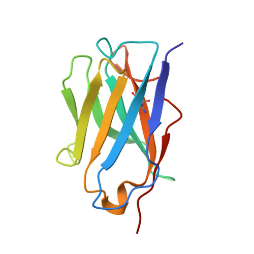
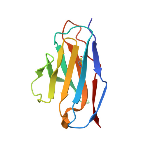
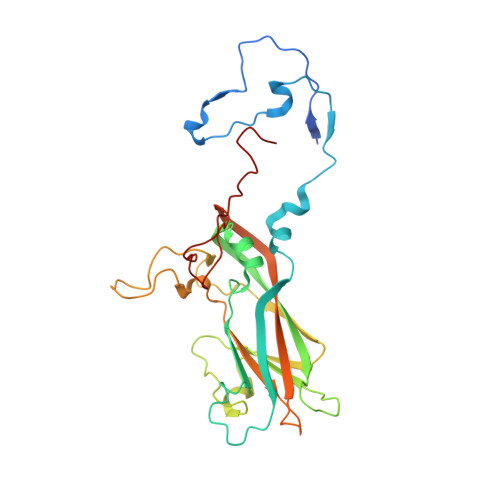
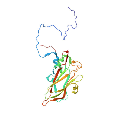
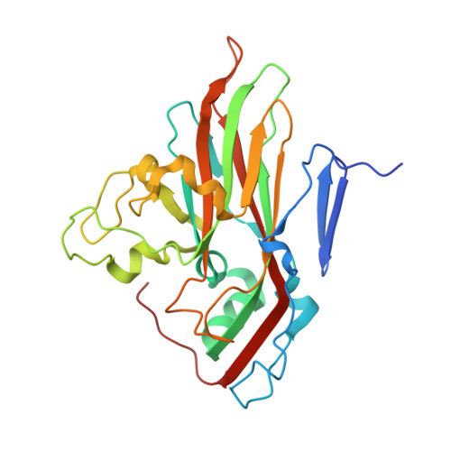
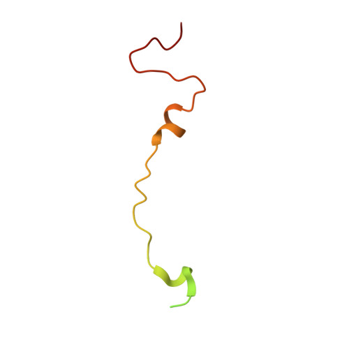
Antibody-induced uncoating of human rhinovirus B14.

Dong, Y.Liu, Y.Jiang, W.Smith, T.J.Xu, Z.Rossmann, M.G.

(2017) Proc Natl Acad Sci U S A 114: 8017-8022

  • DOI: https://doi.org/10.1073/pnas.1707369114
  • Primary Citation Related Structures: 
    5W3E, 5W3L, 5W3M, 5W3O

  • PubMed Abstract: 

    Rhinoviruses (RVs) are the major causes of common colds in humans. They have a nonenveloped, icosahedral capsid surrounding a positive-strand RNA genome. Here we report that the antigen-binding (Fab) fragment of a neutralizing antibody (C5) can trigger genome release from RV-B14 to form emptied particles and neutralize virus infection. Using cryo-electron microscopy, structures of the C5 Fab in complex with the full and emptied particles have been determined at 2.3 Å and 3.0 Å resolution, respectively. Each of the 60 Fab molecules binds primarily to a region on viral protein 3 (VP3). Binding of the C5 Fabs to RV-B14 results in significant conformational changes around holes in the capsid through which the viral RNA might exit. These results are so far the highest resolution view of an antibody-virus complex and elucidate a mechanism whereby antibodies neutralize RVs and related viruses by inducing virus uncoating.


  • Organizational Affiliation
    • Department of Biological Sciences, Purdue University, West Lafayette, IN 47907.

Macromolecule Content 

  • Total Structure Weight: 117.37 kDa 
  • Atom Count: 8,180 
  • Modeled Residue Count: 1,024 
  • Deposited Residue Count: 1,078 
  • Unique protein chains: 6

Macromolecules

Find similar proteins by:|  3D Structure
Entity ID: 1
MoleculeChains  Sequence LengthOrganismDetailsImage
C5 antibody variable heavy domainA [auth E]116Mus musculusMutation(s): 0 
Find similar proteins by:|  3D Structure
Entity ID: 2
MoleculeChains  Sequence LengthOrganismDetailsImage
C5 antibody variable light domainB [auth G]107Mus musculusMutation(s): 0 
Find similar proteins by:|  3D Structure
Entity ID: 3
MoleculeChains  Sequence LengthOrganismDetailsImage
viral protein 1C [auth A]289rhinovirus B14Mutation(s): 0 
UniProt
Find proteins for P03303 (Human rhinovirus 14)
Explore P03303 
Go to UniProtKB:  P03303
Entity Groups
Sequence Clusters30% Identity50% Identity70% Identity90% Identity95% Identity100% Identity
UniProt GroupP03303
Sequence Annotations
Expand
Reference Sequence
Find similar proteins by:|  3D Structure
Entity ID: 4
MoleculeChains  Sequence LengthOrganismDetailsImage
viral protein 3D [auth B]236rhinovirus B14Mutation(s): 0 
UniProt
Find proteins for P03303 (Human rhinovirus 14)
Explore P03303 
Go to UniProtKB:  P03303
Entity Groups
Sequence Clusters30% Identity50% Identity70% Identity90% Identity95% Identity100% Identity
UniProt GroupP03303
Sequence Annotations
Expand
Reference Sequence
Find similar proteins by:|  3D Structure
Entity ID: 5
MoleculeChains  Sequence LengthOrganismDetailsImage
viral protein 2E [auth C]262rhinovirus B14Mutation(s): 0 
UniProt
Find proteins for P03303 (Human rhinovirus 14)
Explore P03303 
Go to UniProtKB:  P03303
Entity Groups
Sequence Clusters30% Identity50% Identity70% Identity90% Identity95% Identity100% Identity
UniProt GroupP03303
Sequence Annotations
Expand
Reference Sequence
Find similar proteins by:|  3D Structure
Entity ID: 6
MoleculeChains  Sequence LengthOrganismDetailsImage
viral protein 4F [auth D]68rhinovirus B14Mutation(s): 0 
UniProt
Find proteins for P03303 (Human rhinovirus 14)
Explore P03303 
Go to UniProtKB:  P03303
Entity Groups
Sequence Clusters30% Identity50% Identity70% Identity90% Identity95% Identity100% Identity
UniProt GroupP03303
Sequence Annotations
Expand
Reference Sequence

Experimental Data & Validation

Experimental Data

  • Method: ELECTRON MICROSCOPY
  • Resolution: 2.26 Å
  • Aggregation State: PARTICLE 
  • Reconstruction Method: SINGLE PARTICLE 
EM Software:
TaskSoftware PackageVersion
MODEL REFINEMENTPHENIX
MODEL REFINEMENTCoot
RECONSTRUCTIONjspr

Structure Validation

View Full Validation Report


Entry History 

& Funding Information

Deposition Data


Funding OrganizationLocationGrant Number
National Institutes of Health/National Institute Of Allergy and Infectious Diseases (NIH/NIAID)United StatesAI011219

Revision History  (Full details and data files)

  • Version 1.0: 2017-07-12
    Type: Initial release
  • Version 1.1: 2017-07-26
    Changes: Author supporting evidence, Database references
  • Version 1.2: 2017-08-09
    Changes: Database references
  • Version 1.3: 2018-07-18
    Changes: Data collection
  • Version 1.4: 2019-12-11
    Changes: Author supporting evidence
  • Version 1.5: 2024-10-30
    Changes: Data collection, Database references, Derived calculations, Structure summary