The crystal structure of Proteus vulgaris tryptophan indole-lyase complexed with oxindolyl-L-alanine: implications for the reaction mechanism.
Phillips, R.S., Buisman, A.A., Choi, S., Hussaini, A., Wood, Z.A.(2018) Acta Crystallogr D Struct Biol 74: 748-759
- PubMed: 30082510 Search on PubMed
- DOI: https://doi.org/10.1107/S2059798318003352
- Primary Citation Related Structures:
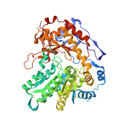
5W19, 5W1B - PubMed Abstract:
Tryptophan indole-lyase (TIL) is a bacterial enzyme which catalyzes the reversible formation of indole and ammonium pyruvate from L-tryptophan. Oxindolyl-L-alanine (OIA) is an inhibitor of TIL, with a K i value of about 5 µM. The crystal structure of the complex of Proteus vulgaris TIL with OIA has now been determined at 2.1 Å resolution. The ligand forms a closed quinonoid complex with the pyridoxal 5'-phosphate (PLP) cofactor. The small domain rotates about 10° to close the active site, bringing His458 into position to donate a hydrogen bond to Asp133, which also accepts a hydrogen bond from the heterocyclic NH of the inhibitor. This brings Phe37 and Phe459 into van der Waals contact with the aromatic ring of OIA. Mutation of the homologous Phe464 in Escherichia coli TIL to Ala results in a 500-fold decrease in k cat /K m for L-tryptophan, with less effect on the reaction of other nonphysiological β-elimination substrates. Stopped-flow kinetic experiments of F464A TIL show that the mutation has no effect on the formation of quinonoid intermediates. An aminoacrylate intermediate is observed in the reaction of F464A TIL with S-ethyl-L-cysteine and benzimidazole. A model of the L-tryptophan quinonoid complex with PLP in the active site of P. vulgaris TIL shows that there would be a severe clash of Phe459 (∼1.5 Å apart) and Phe37 (∼2 Å apart) with the benzene ring of the substrate. It is proposed that this creates distortion of the substrate aromatic ring out of plane and moves the substrate upwards on the reaction coordinate towards the transition state, thus reducing the activation energy and accelerating the enzymatic reaction.
- Department of Chemistry, University of Georgia, Athens, GA 30602, USA.
Organizational Affiliation: