


Crystal structure of glutamate racemase from Thermus Thermophilus in complex with D-glutamate
Cooling, G.T., Vance, N.R., Spies, M.A.To be published.
Experimental Data Snapshot
Starting Model: experimental
View more details
wwPDB Validation 3D Report Full Report

Entity ID: 1 | |||||
|---|---|---|---|---|---|
| Molecule | Chains | Sequence Length | Organism | Details | Image |
| Glutamate racemase | 278 | Thermus thermophilus | Mutation(s): 0 Gene Names: murI, TTHA1643 EC: 5.1.1.3 | 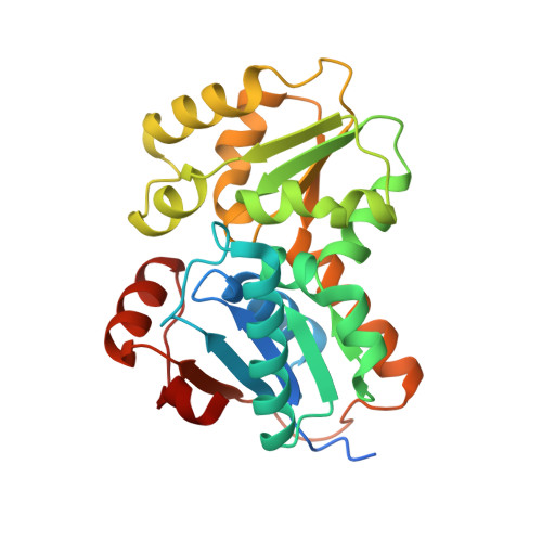 | |
UniProt | |||||
Entity Groups | |||||
| Sequence Clusters | 30% Identity50% Identity70% Identity90% Identity95% Identity100% Identity | ||||
| UniProt Group | Q5SHT7 | ||||
Sequence AnnotationsExpand | |||||
Reference Sequence | |||||
| Ligands 3 Unique | |||||
|---|---|---|---|---|---|
| ID | Chains | Name / Formula / InChI Key | 2D Diagram | 3D Interactions | |
| DGL Download:Ideal Coordinates CCD File | E [auth A], L [auth B], Q [auth C], Z [auth D] | D-GLUTAMIC ACID C5 H9 N O4 WHUUTDBJXJRKMK-GSVOUGTGSA-N |  | ||
| NO3 Download:Ideal Coordinates CCD File | AA [auth D] BA [auth D] CA [auth D] DA [auth D] F [auth A] | NITRATE ION N O3 NHNBFGGVMKEFGY-UHFFFAOYSA-N |  | ||
| CL Download:Ideal Coordinates CCD File | EA [auth D] FA [auth D] K [auth A] O [auth B] P [auth B] | CHLORIDE ION Cl VEXZGXHMUGYJMC-UHFFFAOYSA-M |  | ||
| Length ( Å ) | Angle ( ˚ ) |
|---|---|
| a = 81.677 | α = 90 |
| b = 96.728 | β = 90 |
| c = 179.066 | γ = 90 |
| Software Name | Purpose |
|---|---|
| PHENIX | refinement |
| XDS | data reduction |
| SCALA | data scaling |
| PHASER | phasing |
| Funding Organization | Location | Grant Number |
|---|---|---|
| National Institutes of Health/National Institute of General Medical Sciences (NIH/NIGMS) | United States | R01 GM097373 |


