5VYB | pdb_00005vyb

Structure of the carbohydrate recognition domain of Dectin-2 complexed with a mammalian-type high mannose Man9GlcNAc2 oligosaccharide


Experimental Data Snapshot

  • Method: X-RAY DIFFRACTION
  • Resolution: 2.40 Å
  • R-Value Free: 
    0.240 (Depositor), 0.241 (DCC) 
  • R-Value Work: 
    0.205 (Depositor), 0.209 (DCC) 
  • R-Value Observed: 
    0.207 (Depositor) 

Starting Model: experimental
View more details

wwPDB Validation 3D Report Full Report

Validation slider image for 5VYB

This is version 2.2 of the entry. See complete history

Literature

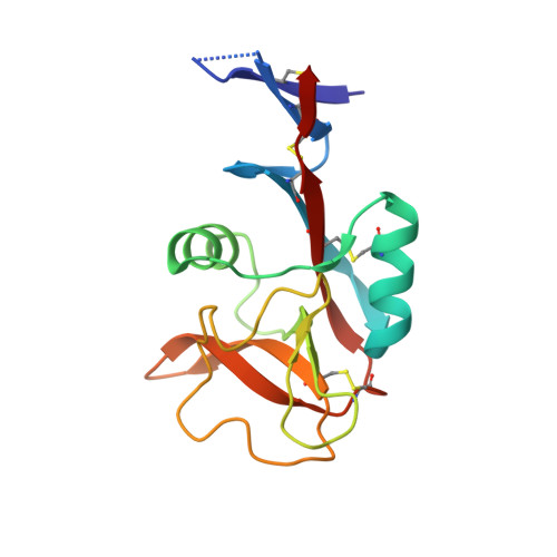
Mechanism of pathogen recognition by human dectin-2.

Feinberg, H.Jegouzo, S.A.F.Rex, M.J.Drickamer, K.Weis, W.I.Taylor, M.E.

(2017) J Biological Chem 292: 13402-13414

  • DOI: https://doi.org/10.1074/jbc.M117.799080
  • Primary Citation Related Structures: 
    5VYB

  • PubMed Abstract: 

    Dectin-2, a C-type lectin on macrophages and other cells of the innate immune system, functions in response to pathogens, particularly fungi. The carbohydrate-recognition domain (CRD) in dectin-2 is linked to a transmembrane sequence that interacts with the common Fc receptor γ subunit to initiate immune signaling. The molecular mechanism by which dectin-2 selectively binds to pathogens has been investigated by characterizing the CRD expressed in a bacterial system. Competition binding studies indicated that the CRD binds to monosaccharides with modest affinity and that affinity was greatly enhanced for mannose-linked α1-2 or α1-4 to a second mannose residue. Glycan array analysis confirmed selective binding of the CRD to glycans that contain Manα1-2Man epitopes. Crystals of the CRD in complex with a mammalian-type high-mannose Man 9 GlcNAc 2 oligosaccharide exhibited interaction with Manα1-2Man on two different termini of the glycan, with the reducing-end mannose residue ligated to Ca 2+ in a primary binding site and the nonreducing terminal mannose residue occupying an adjacent secondary site. Comparison of the binding sites in DC-SIGN and langerin, two other pathogen-binding receptors of the innate immune system, revealed why these two binding sites accommodate only terminal Manα1-2Man structures, whereas dectin-2 can bind Manα1-2Man in internal positions in mannans and other polysaccharides. The specificity and geometry of the dectin-2-binding site provide the molecular mechanism for binding of dectin-2 to fungal mannans and also to bacterial lipopolysaccharides, capsular polysaccharides, and lipoarabinomannans that contain the Manα1-2Man disaccharide unit.


  • Organizational Affiliation
    • From the Departments of Structural Biology and Molecular and Cellular Physiology, Stanford University School of Medicine, Stanford, California 94305 and.

Macromolecule Content 

  • Total Structure Weight: 18.31 kDa 
  • Atom Count: 1,269 
  • Modeled Residue Count: 143 
  • Deposited Residue Count: 147 
  • Unique protein chains: 1

Macromolecules

Find similar proteins by:|  3D Structure
Entity ID: 1
MoleculeChains  Sequence LengthOrganismDetailsImage
C-type lectin domain family 6 member A147Homo sapiensMutation(s): 0 
Gene Names: CLEC6ACLECSF10DECTIN2
UniProt & NIH Common Fund Data Resources
Find proteins for Q6EIG7 (Homo sapiens)
Explore Q6EIG7 
Go to UniProtKB:  Q6EIG7
PHAROS:  Q6EIG7
GTEx:  ENSG00000205846 
Entity Groups
Sequence Clusters30% Identity50% Identity70% Identity90% Identity95% Identity100% Identity
UniProt GroupQ6EIG7
Sequence Annotations
Expand
Reference Sequence

Oligosaccharides

Help  
Entity ID: 2
MoleculeChains Length2D Diagram GlycosylationD Interactions
alpha-D-mannopyranose-(1-2)-alpha-D-mannopyranose-(1-2)-alpha-D-mannopyranose-(1-3)-[alpha-D-mannopyranose-(1-2)-alpha-D-mannopyranose-(1-3)-alpha-D-mannopyranose-(1-6)]beta-D-mannopyranose
B
7N/A
Glycosylation Resources
GlyTouCan: G78528AJ
GlyCosmos: G78528AJ

Experimental Data & Validation

Experimental Data

  • Method: X-RAY DIFFRACTION
  • Resolution: 2.40 Å
  • R-Value Free:  0.240 (Depositor), 0.241 (DCC) 
  • R-Value Work:  0.205 (Depositor), 0.209 (DCC) 
  • R-Value Observed: 0.207 (Depositor) 
Space Group: C 2 2 21
Unit Cell:
Length ( Å )Angle ( ˚ )
a = 64.055α = 90
b = 78.834β = 90
c = 76.459γ = 90
Software Package:
Software NamePurpose
Aimlessdata scaling
PHENIXrefinement
PDB_EXTRACTdata extraction
XDSdata reduction
PHASERphasing

Structure Validation

View Full Validation Report


Entry History 

& Funding Information

Deposition Data


Funding OrganizationLocationGrant Number
Biotechnology and Biological Sciences Research Council (BBSRC)United KingdomBB/K007718/1
Wellcome TrustUnited Kingdom--
National Institutes of Health/National Institute of General Medical Sciences (NIH/NIGMS)United StatesGM62116

Revision History  (Full details and data files)

  • Version 1.0: 2017-07-05
    Type: Initial release
  • Version 1.1: 2017-07-12
    Changes: Database references
  • Version 1.2: 2017-08-23
    Changes: Author supporting evidence, Data collection, Database references
  • Version 1.3: 2018-03-21
    Changes: Derived calculations
  • Version 1.4: 2020-01-01
    Changes: Author supporting evidence
  • Version 2.0: 2020-07-29
    Type: Remediation
    Reason: Carbohydrate remediation
    Changes: Atomic model, Data collection, Derived calculations, Structure summary
  • Version 2.1: 2023-10-04
    Changes: Data collection, Database references, Derived calculations, Refinement description, Structure summary
  • Version 2.2: 2024-11-20
    Changes: Structure summary