5VU7

TNA polymerase, open ternary complex


Experimental Data Snapshot

  • Method: X-RAY DIFFRACTION
  • Resolution: 2.72 Å
  • R-Value Free: 0.268 
  • R-Value Work: 0.214 
  • R-Value Observed: 0.218 

wwPDB Validation   3D Report Full Report


Ligand Structure Quality Assessment 


This is version 1.1 of the entry. See complete history


Literature

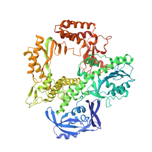
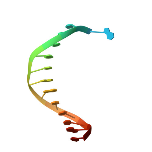
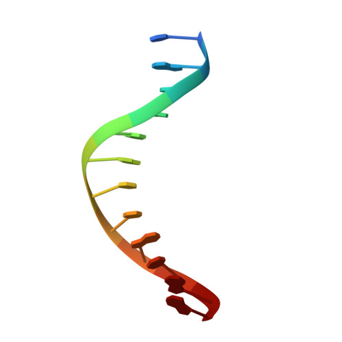
Structural basis for TNA synthesis by an engineered TNA polymerase.

Chim, N.Shi, C.Sau, S.P.Nikoomanzar, A.Chaput, J.C.

(2017) Nat Commun 8: 1810-1810

  • DOI: https://doi.org/10.1038/s41467-017-02014-0
  • Primary Citation of Related Structures:  
    5VU5, 5VU6, 5VU7, 5VU8, 5VU9

  • PubMed Abstract: 

    Darwinian evolution experiments carried out on xeno-nucleic acid (XNA) polymers require engineered polymerases that can faithfully and efficiently copy genetic information back and forth between DNA and XNA. However, current XNA polymerases function with inferior activity relative to their natural counterparts. Here, we report five X-ray crystal structures that illustrate the pathway by which α-(L)-threofuranosyl nucleic acid (TNA) triphosphates are selected and extended in a template-dependent manner using a laboratory-evolved polymerase known as Kod-RI. Structural comparison of the apo, binary, open and closed ternary, and translocated product detail an ensemble of interactions and conformational changes required to promote TNA synthesis. Close inspection of the active site in the closed ternary structure reveals a sub-optimal binding geometry that explains the slow rate of catalysis. This key piece of information, which is missing for all naturally occurring archaeal DNA polymerases, provides a framework for engineering new TNA polymerase variants.


  • Organizational Affiliation

    Departments of Pharmaceutical Sciences, Chemistry, and Molecular Biology and Biochemistry University of California, Irvine, CA, 92697-3958, USA.


Macromolecules

Find similar proteins by:  (by identity cutoff)  |  3D Structure
Entity ID: 1
MoleculeChains Sequence LengthOrganismDetailsImage
DNA polymerase774Thermococcus kodakarensisMutation(s): 6 
EC: 2.7.7.7
UniProt
Find proteins for D0VWU9 (Thermococcus kodakarensis (strain ATCC BAA-918 / JCM 12380 / KOD1))
Explore D0VWU9 
Go to UniProtKB:  D0VWU9
Entity Groups  
Sequence Clusters30% Identity50% Identity70% Identity90% Identity95% Identity100% Identity
UniProt GroupD0VWU9
Sequence Annotations
Expand
  • Reference Sequence

Find similar nucleic acids by:  Sequence   |   3D Structure  

Entity ID: 2
MoleculeChains LengthOrganismImage
DNA templateB [auth T]16synthetic construct
Sequence Annotations
Expand
  • Reference Sequence

Find similar nucleic acids by:  Sequence   |   3D Structure  

Entity ID: 3
MoleculeChains LengthOrganismImage
DNA/TNA hybrid primerC [auth P]12synthetic construct
Sequence Annotations
Expand
  • Reference Sequence
Small Molecules
Ligands 3 Unique
IDChains Name / Formula / InChI Key2D Diagram3D Interactions
FA2
Query on FA2

Download Ideal Coordinates CCD File 
F [auth A]5-(6-AMINO-9H-PURIN-9-YL)-4-HYDROXYTETRAHYDROFURAN-3-YL DIHYDROGEN PHOSPHATE
C9 H12 N5 O6 P
IJEJRDCFMFEDGL-AFEQZKEHSA-N
SO4
Query on SO4

Download Ideal Coordinates CCD File 
E [auth A]SULFATE ION
O4 S
QAOWNCQODCNURD-UHFFFAOYSA-L
MG
Query on MG

Download Ideal Coordinates CCD File 
D [auth A]MAGNESIUM ION
Mg
JLVVSXFLKOJNIY-UHFFFAOYSA-N
Experimental Data & Validation

Experimental Data

  • Method: X-RAY DIFFRACTION
  • Resolution: 2.72 Å
  • R-Value Free: 0.268 
  • R-Value Work: 0.214 
  • R-Value Observed: 0.218 
  • Space Group: P 2 21 21
Unit Cell:
Length ( Å )Angle ( ˚ )
a = 66.55α = 90
b = 112.61β = 90
c = 145.46γ = 90
Software Package:
Software NamePurpose
PHENIXrefinement
iMOSFLMdata reduction
Aimlessdata scaling
PHASERphasing

Structure Validation

View Full Validation Report


Ligand Structure Quality Assessment 


Entry History 

Deposition Data

Revision History  (Full details and data files)

  • Version 1.0: 2017-12-06
    Type: Initial release
  • Version 1.1: 2017-12-13
    Changes: Database references