5VSG | pdb_00005vsg

Fibrils of the super helical repeat peptide, SHR-FF, grown at elevated temperature


Experimental Data Snapshot

  • Method: X-RAY DIFFRACTION
  • Resolution: 1.10 Å
  • R-Value Free: 
    0.134 (Depositor), 0.133 (DCC) 
  • R-Value Work: 
    0.114 (Depositor), 0.113 (DCC) 
  • R-Value Observed: 
    0.117 (Depositor) 

wwPDB Validation 3D Report Full Report

Validation slider image for 5VSG

This is version 1.3 of the entry. See complete history

Literature

Transition of Metastable Cross-alpha Crystals into Cross-beta Fibrils by beta-Turn Flipping.

Mondal, S.Jacoby, G.Sawaya, M.R.Arnon, Z.A.Adler-Abramovich, L.Rehak, P.Vukovic, L.Shimon, L.J.W.Kral, P.Beck, R.Gazit, E.

(2019) J Am Chem Soc 141: 363-369

  • DOI: https://doi.org/10.1021/jacs.8b10289
  • Primary Citation Related Structures: 
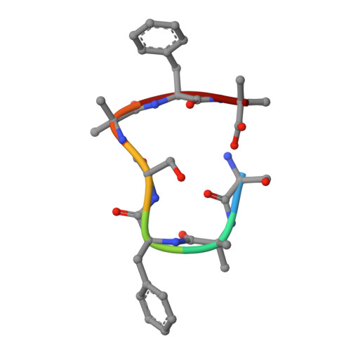
    5VSG

  • PubMed Abstract: 

    The ensemble of native, folded state was once considered to represent the global energy minimum of a given protein sequence. More recently, the discovery of the cross-β amyloid state revealed that deeper energy minima exist, often associated with pathogenic, fibrillar deposits, when the concentration of proteins reaches a critical value. Fortunately, a sizable energy barrier impedes the conversion from native to pathogenic states. However, little is known about the structure of the related transition state. In addition, there are indications of polymorphism in the amyloidogenic process. Here, we report the first evidence of the conversion of metastable cross-α-helical crystals to thermodynamically stable cross-β-sheet-like fibrils by a de novo designed heptapeptide. Furthermore, for the first time, we demonstrate at atomic resolution that the flip of a peptide plane from a type I to a type II' turn facilitates transformation to cross-β structure and assembly of a dry steric zipper. This study establishes the potential of a peptide turn, a common protein secondary structure, to serve as a principal gatekeeper between a native metastable folded state and the amyloid state.


  • Organizational Affiliation
    • Department of Molecular Microbiology and Biotechnology, George S. Wise Faculty of Life Sciences , Tel Aviv University , Tel Aviv 69978 , Israel.

Macromolecule Content 

  • Total Structure Weight: 0.74 kDa 
  • Atom Count: 55 
  • Modeled Residue Count: 7 
  • Deposited Residue Count: 7 
  • Unique protein chains: 1

Macromolecules

Find similar proteins by:  Sequence   |   3D Structure  
Entity ID: 1
MoleculeChains  Sequence LengthOrganismDetailsImage
Super Helical Repeat Peptide SHR-FF7synthetic constructMutation(s): 0 

Small Molecules

Modified Residues  1 Unique
IDChains TypeFormula2D DiagramParent
AIB
Query on AIB
A
L-PEPTIDE LINKINGC4 H9 N O2ALA

Experimental Data & Validation

Experimental Data

  • Method: X-RAY DIFFRACTION
  • Resolution: 1.10 Å
  • R-Value Free:  0.134 (Depositor), 0.133 (DCC) 
  • R-Value Work:  0.114 (Depositor), 0.113 (DCC) 
  • R-Value Observed: 0.117 (Depositor) 
Space Group: P 1 21 1
Unit Cell:
Length ( Å )Angle ( ˚ )
a = 5.343α = 90
b = 20.883β = 93.9
c = 16.795γ = 90
Software Package:
Software NamePurpose
DENZOdata collection
SCALEPACKdata scaling
REFMACrefinement
PDB_EXTRACTdata extraction
DENZOdata reduction
SHELXDphasing

Structure Validation

View Full Validation Report


Entry History 

& Funding Information

Deposition Data


Funding OrganizationLocationGrant Number
National Institutes of Health/Office of the DirectorUnited StatesS10OD021527
National Institutes of Health/National Institute of General Medical Sciences (NIH/NIGMS)United StatesP41 GM103403

Revision History  (Full details and data files)

  • Version 1.0: 2018-06-27
    Type: Initial release
  • Version 1.1: 2019-07-10
    Changes: Data collection, Database references
  • Version 1.2: 2020-01-01
    Changes: Author supporting evidence
  • Version 1.3: 2025-04-02
    Changes: Data collection, Database references, Structure summary