5VPN | pdb_00005vpn

E. coli Quinol fumarate reductase FrdA E245Q mutation


Experimental Data Snapshot

  • Method: X-RAY DIFFRACTION
  • Resolution: 4.22 Å
  • R-Value Free: 
    0.270 (Depositor), 0.277 (DCC) 
  • R-Value Work: 
    0.215 (Depositor), 0.221 (DCC) 
  • R-Value Observed: 
    0.218 (Depositor) 

Starting Model: experimental
View more details

wwPDB Validation 3D Report Full Report

Validation slider image for 5VPN

Ligand Structure Quality Assessment 


This is version 1.5 of the entry. See complete history

Literature

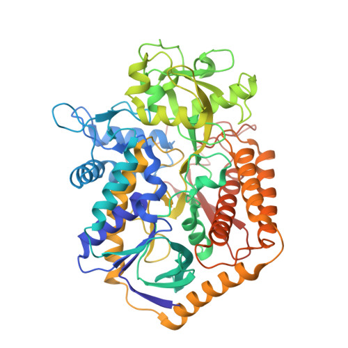
Structural and biochemical analyses reveal insights into covalent flavinylation of the Escherichia coli Complex II homolog quinol:fumarate reductase.

Starbird, C.A.Maklashina, E.Sharma, P.Qualls-Histed, S.Cecchini, G.Iverson, T.M.

(2017) J Biological Chem 292: 12921-12933

  • DOI: https://doi.org/10.1074/jbc.M117.795120
  • Primary Citation Related Structures: 
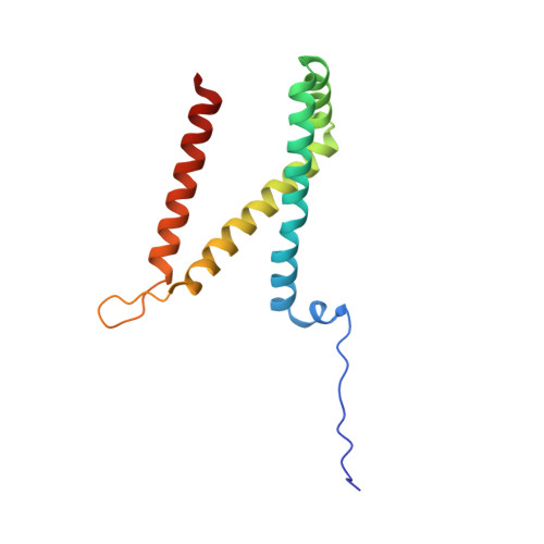
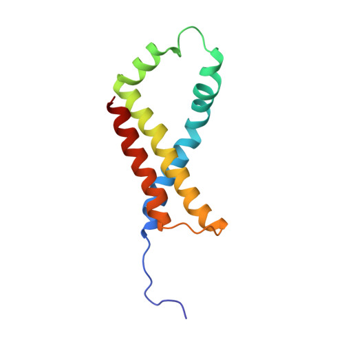
    5VPN

  • PubMed Abstract: 

    The Escherichia coli Complex II homolog quinol:fumarate reductase (QFR, FrdABCD) catalyzes the interconversion of fumarate and succinate at a covalently attached FAD within the FrdA subunit. The SdhE assembly factor enhances covalent flavinylation of Complex II homologs, but the mechanisms underlying the covalent attachment of FAD remain to be fully elucidated. Here, we explored the mechanisms of covalent flavinylation of the E. coli QFR FrdA subunit. Using a Δ sdhE E. coli strain, we show that the requirement for the assembly factor depends on the cellular redox environment. We next identified residues important for the covalent attachment and selected the FrdA E245 residue, which contributes to proton shuttling during fumarate reduction, for detailed biophysical and structural characterization. We found that QFR complexes containing FrdA E245Q have a structure similar to that of the WT flavoprotein, but lack detectable substrate binding and turnover. In the context of the isolated FrdA subunit, the anticipated assembly intermediate during covalent flavinylation, FrdA E245 variants had stability similar to that of WT FrdA, contained noncovalent FAD, and displayed a reduced capacity to interact with SdhE. However, small-angle X-ray scattering (SAXS) analysis of WT FrdA cross-linked to SdhE suggested that the FrdA E245 residue is unlikely to contribute directly to the FrdA-SdhE protein-protein interface. We also found that no auxiliary factor is absolutely required for flavinylation, indicating that the covalent flavinylation is autocatalytic. We propose that multiple factors, including the SdhE assembly factor and bound dicarboxylates, stimulate covalent flavinylation by preorganizing the active site to stabilize the quinone-methide intermediate.


  • Organizational Affiliation
    • Graduate Program in Chemical and Physical Biology, Vanderbilt University, Nashville, Tennessee 37232.

Macromolecule Content 

  • Total Structure Weight: 241.98 kDa 
  • Atom Count: 16,845 
  • Modeled Residue Count: 2,146 
  • Deposited Residue Count: 2,154 
  • Unique protein chains: 4

Macromolecules

Find similar proteins by:|  3D Structure
Entity ID: 1
MoleculeChains  Sequence LengthOrganismDetailsImage
Fumarate reductase flavoprotein subunit
A, E
585Escherichia coli K-12Mutation(s): 1 
Gene Names: frdAb4154JW4115
EC: 1.3.5.4 (PDB Primary Data), 1.3.5.1 (UniProt)
Membrane Entity: Yes 
UniProt
Find proteins for P00363 (Escherichia coli (strain K12))
Explore P00363 
Go to UniProtKB:  P00363
Entity Groups
Sequence Clusters30% Identity50% Identity70% Identity90% Identity95% Identity100% Identity
UniProt GroupP00363
Sequence Annotations
Expand
Reference Sequence
Find similar proteins by:|  3D Structure
Entity ID: 2
MoleculeChains  Sequence LengthOrganismDetailsImage
Succinate dehydrogenase iron-sulfur subunit
B, F
243Escherichia coliMutation(s): 0 
EC: 1.3.5.1
Membrane Entity: Yes 
UniProt
Find proteins for P0AC47 (Escherichia coli (strain K12))
Explore P0AC47 
Go to UniProtKB:  P0AC47
Entity Groups
Sequence Clusters30% Identity50% Identity70% Identity90% Identity95% Identity100% Identity
UniProt GroupP0AC47
Sequence Annotations
Expand
Reference Sequence
Find similar proteins by:|  3D Structure
Entity ID: 3
MoleculeChains  Sequence LengthOrganismDetailsImage
Fumarate reductase subunit C
C, G
130Escherichia coli K-12Mutation(s): 0 
Gene Names: frdCb4152JW4113
Membrane Entity: Yes 
UniProt
Find proteins for P0A8Q0 (Escherichia coli (strain K12))
Explore P0A8Q0 
Go to UniProtKB:  P0A8Q0
Entity Groups
Sequence Clusters30% Identity50% Identity70% Identity90% Identity95% Identity100% Identity
UniProt GroupP0A8Q0
Sequence Annotations
Expand
Reference Sequence
Find similar proteins by:|  3D Structure
Entity ID: 4
MoleculeChains  Sequence LengthOrganismDetailsImage
Fumarate reductase subunit D
D, H
119Escherichia coli K-12Mutation(s): 0 
Gene Names: frdDb4151JW4112
Membrane Entity: Yes 
UniProt
Find proteins for P0A8Q3 (Escherichia coli (strain K12))
Explore P0A8Q3 
Go to UniProtKB:  P0A8Q3
Entity Groups
Sequence Clusters30% Identity50% Identity70% Identity90% Identity95% Identity100% Identity
UniProt GroupP0A8Q3
Sequence Annotations
Expand
Reference Sequence

Small Molecules

Ligands 4 Unique
IDChains Name / Formula / InChI Key2D Diagram3D Interactions
FAD

Query on FAD


Download:Ideal Coordinates CCD File
I [auth A],
M [auth E]
FLAVIN-ADENINE DINUCLEOTIDE
C27 H33 N9 O15 P2
VWWQXMAJTJZDQX-UYBVJOGSSA-N
SF4

Query on SF4


Download:Ideal Coordinates CCD File
L [auth B],
P [auth F]
IRON/SULFUR CLUSTER
Fe4 S4
LJBDFODJNLIPKO-UHFFFAOYSA-N
F3S

Query on F3S


Download:Ideal Coordinates CCD File
K [auth B],
O [auth F]
FE3-S4 CLUSTER
Fe3 S4
FCXHZBQOKRZXKS-UHFFFAOYSA-N
FES

Query on FES


Download:Ideal Coordinates CCD File
J [auth B],
N [auth F]
FE2/S2 (INORGANIC) CLUSTER
Fe2 S2
NIXDOXVAJZFRNF-UHFFFAOYSA-N

Experimental Data & Validation

Experimental Data

  • Method: X-RAY DIFFRACTION
  • Resolution: 4.22 Å
  • R-Value Free:  0.270 (Depositor), 0.277 (DCC) 
  • R-Value Work:  0.215 (Depositor), 0.221 (DCC) 
  • R-Value Observed: 0.218 (Depositor) 
Space Group: P 21 21 21
Unit Cell:
Length ( Å )Angle ( ˚ )
a = 133.565α = 90
b = 138.134β = 90
c = 220.079γ = 90
Software Package:
Software NamePurpose
PHENIXrefinement
PDB_EXTRACTdata extraction
HKL-2000data scaling
PHASERphasing

Structure Validation

View Full Validation Report


Ligand Structure Quality Assessment 


Entry History 

& Funding Information

Deposition Data


Funding OrganizationLocationGrant Number
Department of Veterans AffairsUnited StatesBX001077
National Institutes of Health/National Institute of General Medical Sciences (NIH/NIGMS)United StatesGM061606
National Institutes of Health/National Institute of General Medical Sciences (NIH/NIGMS)United StatesT32GM008320
National Science Foundation (NSF, United States)United StatesDGE:0909667
National Institutes of Health/National Institute of General Medical Sciences (NIH/NIGMS)United StatesR01GM105404
High-End Instrumentation GrantUnited StatesS10OD018483

Revision History  (Full details and data files)

  • Version 1.0: 2017-06-21
    Type: Initial release
  • Version 1.1: 2017-06-28
    Changes: Database references
  • Version 1.2: 2017-08-16
    Changes: Database references
  • Version 1.3: 2017-09-06
    Changes: Author supporting evidence
  • Version 1.4: 2019-11-27
    Changes: Author supporting evidence
  • Version 1.5: 2023-10-04
    Changes: Data collection, Database references, Refinement description, Source and taxonomy, Structure summary