


Crystal structure of 3-oxoacyl-[acyl-carrier protein] reductase from Brucella melitensis in complex with NAD
Mayclin, S.J., Abendroth, J., Lorimer, D.D., Edwards, T.E.To be published.
Experimental Data Snapshot
Starting Model: experimental
View more details
Entity ID: 1 | |||||
|---|---|---|---|---|---|
| Molecule | Chains | Sequence Length | Organism | Details | Image |
| 3-oxoacyl-(Acyl-carrier protein) reductase | 262 | Brucella melitensis bv. 1 str. 16M | Mutation(s): 0 Gene Names: BMEI0026 EC: 1.1.1.100 | 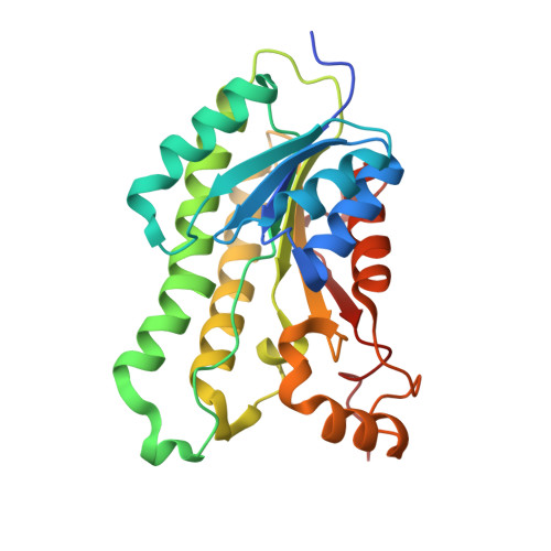 | |
UniProt | |||||
Entity Groups | |||||
| Sequence Clusters | 30% Identity50% Identity70% Identity90% Identity95% Identity100% Identity | ||||
| UniProt Group | Q8YJQ6 | ||||
Sequence AnnotationsExpand | |||||
Reference Sequence | |||||
| Ligands 3 Unique | |||||
|---|---|---|---|---|---|
| ID | Chains | Name / Formula / InChI Key | 2D Diagram | 3D Interactions | |
| NAD Download:Ideal Coordinates CCD File | E [auth A], H [auth B], J [auth C], L [auth D] | NICOTINAMIDE-ADENINE-DINUCLEOTIDE C21 H27 N7 O14 P2 BAWFJGJZGIEFAR-NNYOXOHSSA-N |  | ||
| EDO Download:Ideal Coordinates CCD File | G [auth A] | 1,2-ETHANEDIOL C2 H6 O2 LYCAIKOWRPUZTN-UHFFFAOYSA-N |  | ||
| ACT Download:Ideal Coordinates CCD File | F [auth A], I [auth B], K [auth C], M [auth D] | ACETATE ION C2 H3 O2 QTBSBXVTEAMEQO-UHFFFAOYSA-M |  | ||
| Length ( Å ) | Angle ( ˚ ) |
|---|---|
| a = 74.56 | α = 90 |
| b = 92.71 | β = 90 |
| c = 129.91 | γ = 90 |
| Software Name | Purpose |
|---|---|
| XSCALE | data scaling |
| Coot | model building |
| PHENIX | refinement |
| PDB_EXTRACT | data extraction |
| XDS | data reduction |
| MOLREP | phasing |


