5VLB | pdb_00005vlb

Crystal Structure of Medicago truncatula L-Histidinol Dehydrogenase in Complex with Imidazole


Experimental Data Snapshot

  • Method: X-RAY DIFFRACTION
  • Resolution: 2.25 Å
  • R-Value Free: 
    0.234 (Depositor), 0.236 (DCC) 
  • R-Value Work: 
    0.181 (Depositor), 0.186 (DCC) 
  • R-Value Observed: 
    0.182 (Depositor) 

Starting Model: experimental
View more details

wwPDB Validation 3D Report Full Report

Validation slider image for 5VLB

This is version 1.2 of the entry. See complete history

Literature

Structures of Medicago truncatula L-Histidinol Dehydrogenase Show Rearrangements Required for NAD(+) Binding and the Cofactor Positioned to Accept a Hydride.

Ruszkowski, M.Dauter, Z.

(2017) Sci Rep 7: 10476-10476

  • DOI: https://doi.org/10.1038/s41598-017-10859-0
  • Primary Citation Related Structures: 
    5VLB, 5VLC, 5VLD

  • PubMed Abstract: 

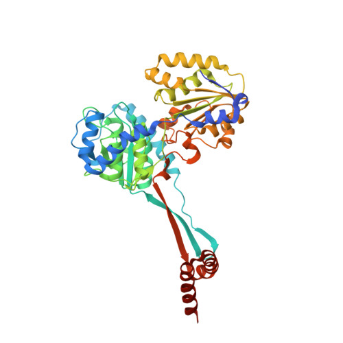
    Plants, lower eukaryotes, bacteria, and archaebacteria synthesise L-histidine (His) in a similar, multistep pathway that is absent in mammals. This makes the His biosynthetic route a promising target for herbicides, antifungal agents, and antibiotics. The last enzyme of the pathway, bifunctional L-histidinol dehydrogenase (HDH, EC 1.1.1.23), catalyses two oxidation reactions: from L-histidinol (HOL) to L-histidinaldehyde and from L-histidinaldehyde to His. Over the course of the reaction, HDH utilises two molecules of NAD + as the hydride acceptor. The object of this study was the HDH enzyme from the model legume plant, Medicago truncatula (MtHDH). Three crystal structures complexed with imidazole, HOL, and His with NAD + provided in-depth insights into the enzyme architecture, its active site, and the cofactor binding mode. The overall structure of MtHDH is similar to the two bacterial orthologues whose three-dimensional structures have been determined. The three snapshots, with the MtHDH enzyme captured in different states, visualise structural rearrangements that allow for NAD + binding for the first time. Furthermore, the MtHDH complex with His and NAD + displays the cofactor molecule situated in a way that would allow for a hydride transfer.


  • Organizational Affiliation
    • Synchrotron Radiation Research Section of MCL, National Cancer Institute, Argonne, IL, USA. milosz.ruszkowski@nih.gov.

Macromolecule Content 

  • Total Structure Weight: 290.61 kDa 
  • Atom Count: 20,467 
  • Modeled Residue Count: 2,598 
  • Deposited Residue Count: 2,676 
  • Unique protein chains: 1

Macromolecules

Find similar proteins by:|  3D Structure
Entity ID: 1
MoleculeChains  Sequence LengthOrganismDetailsImage
Histidinol dehydrogenase, chloroplastic
A, B, C, D, E
A, B, C, D, E, F
446Medicago truncatulaMutation(s): 0 
Gene Names: MTR_2g030520
EC: 1.1.1.23
UniProt
Find proteins for G7IKX3 (Medicago truncatula)
Explore G7IKX3 
Go to UniProtKB:  G7IKX3
Entity Groups
Sequence Clusters30% Identity50% Identity70% Identity90% Identity95% Identity100% Identity
UniProt GroupG7IKX3
Sequence Annotations
Expand
Reference Sequence

Small Molecules

Ligands 3 Unique
IDChains Name / Formula / InChI Key2D Diagram3D Interactions
PEG

Query on PEG


Download:Ideal Coordinates CCD File
I [auth A]
L [auth B]
O [auth C]
R [auth D]
U [auth E]
I [auth A],
L [auth B],
O [auth C],
R [auth D],
U [auth E],
X [auth F]
DI(HYDROXYETHYL)ETHER
C4 H10 O3
MTHSVFCYNBDYFN-UHFFFAOYSA-N
IMD

Query on IMD


Download:Ideal Coordinates CCD File
H [auth A]
K [auth A]
N [auth C]
Q [auth C]
T [auth E]
H [auth A],
K [auth A],
N [auth C],
Q [auth C],
T [auth E],
W [auth E]
IMIDAZOLE
C3 H5 N2
RAXXELZNTBOGNW-UHFFFAOYSA-O
ZN

Query on ZN


Download:Ideal Coordinates CCD File
G [auth A]
J [auth A]
M [auth C]
P [auth C]
S [auth E]
G [auth A],
J [auth A],
M [auth C],
P [auth C],
S [auth E],
V [auth E]
ZINC ION
Zn
PTFCDOFLOPIGGS-UHFFFAOYSA-N

Experimental Data & Validation

Experimental Data

  • Method: X-RAY DIFFRACTION
  • Resolution: 2.25 Å
  • R-Value Free:  0.234 (Depositor), 0.236 (DCC) 
  • R-Value Work:  0.181 (Depositor), 0.186 (DCC) 
  • R-Value Observed: 0.182 (Depositor) 
Space Group: P 1 21 1
Unit Cell:
Length ( Å )Angle ( ˚ )
a = 105.752α = 90
b = 142.814β = 120.16
c = 105.357γ = 90
Software Package:
Software NamePurpose
REFMACrefinement
XDSdata reduction
XDSdata scaling
BALBESphasing

Structure Validation

View Full Validation Report


Entry History 

Deposition Data

Revision History  (Full details and data files)

  • Version 1.0: 2017-09-20
    Type: Initial release
  • Version 1.1: 2018-04-11
    Changes: Data collection, Structure summary
  • Version 1.2: 2023-10-04
    Changes: Data collection, Database references, Derived calculations, Refinement description