5VKS | pdb_00005vks

Crystal structure of P[19] rotavirus VP8* complexed with LNFPI


Experimental Data Snapshot

  • Method: X-RAY DIFFRACTION
  • Resolution: 1.94 Å
  • R-Value Free: 
    0.230 (Depositor), 0.225 (DCC) 
  • R-Value Work: 
    0.201 (Depositor) 
  • R-Value Observed: 
    0.203 (Depositor) 

Starting Model: experimental
View more details

wwPDB Validation 3D Report Full Report

Validation slider image for 5VKS

This is version 2.1 of the entry. See complete history

Literature

Structural basis of glycan specificity of P[19] VP8*: Implications for rotavirus zoonosis and evolution.

Liu, Y.Xu, S.Woodruff, A.L.Xia, M.Tan, M.Kennedy, M.A.Jiang, X.

(2017) PLoS Pathog 13: e1006707-e1006707

  • DOI: https://doi.org/10.1371/journal.ppat.1006707
  • Primary Citation Related Structures: 
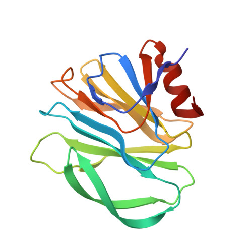
    5VKI, 5VKS

  • PubMed Abstract: 

    Recognition of specific cell surface glycans, mediated by the VP8* domain of the spike protein VP4, is the essential first step in rotavirus (RV) infection. Due to lack of direct structural information of virus-ligand interactions, the molecular basis of ligand-controlled host ranges of the major human RVs (P[8] and P[4]) in P[II] genogroup remains unknown. Here, through characterization of a minor P[II] RV (P[19]) that can infect both animals (pigs) and humans, we made an important advance to fill this knowledge gap by solving the crystal structures of the P[19] VP8* in complex with its ligands. Our data showed that P[19] RVs use a novel binding site that differs from the known ones of other genotypes/genogroups. This binding site is capable of interacting with two types of glycans, the mucin core and type 1 histo-blood group antigens (HBGAs) with a common GlcNAc as the central binding saccharide. The binding site is apparently shared by other P[II] RVs and possibly two genotypes (P[10] and P[12]) in P[I] as shown by their highly conserved GlcNAc-interacting residues. These data provide strong evidence of evolutionary connections among these human and animal RVs, pointing to a common ancestor in P[I] with a possible animal host origin. While the binding properties to GlcNAc-containing saccharides are maintained, changes in binding to additional residues, such as those in the polymorphic type 1 HBGAs may occur in the course of RV evolution, explaining the complex P[II] genogroup that mainly causes diseases in humans but also in some animals.


  • Organizational Affiliation
    • Tianjin Key Laboratory of Molecular Nuclear Medicine, Institute of Radiation Medicine, Chinese Academy of Medical Sciences and Peking Union Medical College, Tianjin, China.

Macromolecule Content 

  • Total Structure Weight: 38.6 kDa 
  • Atom Count: 2,910 
  • Modeled Residue Count: 320 
  • Deposited Residue Count: 320 
  • Unique protein chains: 1

Macromolecules

Find similar proteins by:|  3D Structure
Entity ID: 1
MoleculeChains  Sequence LengthOrganismDetailsImage
Outer capsid protein VP4
A, B
160Human rotavirus AMutation(s): 0 
Gene Names: VP4
UniProt
Find proteins for A7YCM0 (Human rotavirus A)
Explore A7YCM0 
Go to UniProtKB:  A7YCM0
Entity Groups
Sequence Clusters30% Identity50% Identity70% Identity90% Identity95% Identity100% Identity
UniProt GroupA7YCM0
Sequence Annotations
Expand
Reference Sequence

Oligosaccharides

Help  
Entity ID: 2
MoleculeChains Length2D Diagram GlycosylationD Interactions
alpha-L-fucopyranose-(1-2)-beta-D-galactopyranose-(1-3)-2-acetamido-2-deoxy-beta-D-glucopyranose-(1-3)-beta-D-galactopyranose-(1-4)-alpha-D-glucopyranose
C, D
5N/A
Glycosylation Resources
GlyTouCan: G57039PS
GlyCosmos: G57039PS
GlyGen: G57039PS

Experimental Data & Validation

Experimental Data

  • Method: X-RAY DIFFRACTION
  • Resolution: 1.94 Å
  • R-Value Free:  0.230 (Depositor), 0.225 (DCC) 
  • R-Value Work:  0.201 (Depositor) 
  • R-Value Observed: 0.203 (Depositor) 
Space Group: I 41
Unit Cell:
Length ( Å )Angle ( ˚ )
a = 115.631α = 90
b = 115.631β = 90
c = 101.828γ = 90
Software Package:
Software NamePurpose
REFMACrefinement
SCALAdata scaling
PDB_EXTRACTdata extraction
iMOSFLMdata reduction
PHASERphasing

Structure Validation

View Full Validation Report


Entry History 

& Funding Information

Deposition Data


Funding OrganizationLocationGrant Number
National Institutes of Health/National Institute Of Allergy and Infectious Diseases (NIH/NIAID)United States1R56AI11483-01A1

Revision History  (Full details and data files)

  • Version 1.0: 2017-11-08
    Type: Initial release
  • Version 1.1: 2017-12-06
    Changes: Database references
  • Version 1.2: 2019-12-11
    Changes: Author supporting evidence, Data collection
  • Version 2.0: 2020-07-29
    Type: Remediation
    Reason: Carbohydrate remediation
    Changes: Atomic model, Data collection, Derived calculations, Refinement description, Structure summary
  • Version 2.1: 2023-10-04
    Changes: Data collection, Database references, Derived calculations, Refinement description, Structure summary