5VI4 | pdb_00005vi4

IL-33/ST2/IL-1RAcP ternary complex structure


Experimental Data Snapshot

  • Method: X-RAY DIFFRACTION
  • Resolution: 2.79 Å
  • R-Value Free: 
    0.267 (Depositor), 0.270 (DCC) 
  • R-Value Work: 
    0.212 (Depositor), 0.214 (DCC) 
  • R-Value Observed: 
    0.215 (Depositor) 

Starting Models: experimental
View more details

wwPDB Validation 3D Report Full Report

Validation slider image for 5VI4

Ligand Structure Quality Assessment 


This is version 1.4 of the entry. See complete history

Literature

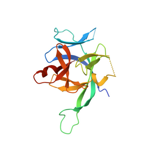
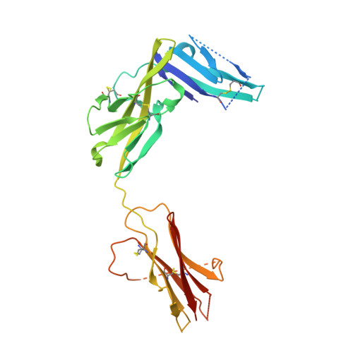
IL-1 Family Cytokines Use Distinct Molecular Mechanisms to Signal through Their Shared Co-receptor.

Gunther, S.Deredge, D.Bowers, A.L.Luchini, A.Bonsor, D.A.Beadenkopf, R.Liotta, L.Wintrode, P.L.Sundberg, E.J.

(2017) Immunity 47: 510-523.e4

  • DOI: https://doi.org/10.1016/j.immuni.2017.08.004
  • Primary Citation Related Structures: 
    5VI4

  • PubMed Abstract: 

    Within the interleukin 1 (IL-1) cytokine family, IL-1 receptor accessory protein (IL-1RAcP) is the co-receptor for eight receptor-cytokine pairs, including those involving cytokines IL-1β and IL-33. Unlike IL-1β, IL-33 does not have a signaling complex that includes both its cognate receptor, ST2, and the shared co-receptor IL-1RAcP, which we now present here. Although the IL-1β and IL-33 complexes shared structural features and engaged identical molecular surfaces of IL-1RAcP, these cytokines had starkly different strategies for co-receptor engagement and signal activation. Our data suggest that IL-1β binds to IL-1RI to properly present the cytokine to IL-1RAcP, whereas IL-33 binds to ST2 in order to conformationally constrain the cognate receptor in an IL-1RAcP-receptive state. These findings indicate that members of the IL-1 family of cytokines use distinct molecular mechanisms to signal through their shared co-receptor, and they provide the foundation from which to design new therapies to target IL-33 signaling.


  • Organizational Affiliation
    • Institute of Human Virology, University of Maryland School of Medicine, Baltimore, MD 21201, USA.

Macromolecule Content 

  • Total Structure Weight: 185.66 kDa 
  • Atom Count: 11,304 
  • Modeled Residue Count: 1,405 
  • Deposited Residue Count: 1,612 
  • Unique protein chains: 3

Macromolecules

Find similar proteins by:|  3D Structure
Entity ID: 1
MoleculeChains  Sequence LengthOrganismDetailsImage
Interleukin-33
A, D
158Mus musculusMutation(s): 4 
Gene Names: Il33
UniProt & NIH Common Fund Data Resources
Find proteins for Q8BVZ5 (Mus musculus)
Explore Q8BVZ5 
Go to UniProtKB:  Q8BVZ5
IMPC:  MGI:1924375
Entity Groups
Sequence Clusters30% Identity50% Identity70% Identity90% Identity95% Identity100% Identity
UniProt GroupQ8BVZ5
Sequence Annotations
Expand
Reference Sequence
Find similar proteins by:|  3D Structure
Entity ID: 2
MoleculeChains  Sequence LengthOrganismDetailsImage
Interleukin-1 receptor-like 1
B, E
309Mus musculusMutation(s): 0 
Gene Names: Il1rl1Ly84St2Ste2
EC: 3.2.2.6
UniProt
Find proteins for P14719 (Mus musculus)
Explore P14719 
Go to UniProtKB:  P14719
Entity Groups
Sequence Clusters30% Identity50% Identity70% Identity90% Identity95% Identity100% Identity
UniProt GroupP14719
Glycosylation
Glycosylation Sites: 1Go to GlyGen: P14719-1
Sequence Annotations
Expand
Reference Sequence
Find similar proteins by:|  3D Structure
Entity ID: 3
MoleculeChains  Sequence LengthOrganismDetailsImage
Interleukin-1 receptor accessory protein
C, F
339Mus musculusMutation(s): 4 
Gene Names: Il1rap
EC: 3.2.2.6
UniProt
Find proteins for Q61730 (Mus musculus)
Explore Q61730 
Go to UniProtKB:  Q61730
Entity Groups
Sequence Clusters30% Identity50% Identity70% Identity90% Identity95% Identity100% Identity
UniProt GroupQ61730
Sequence Annotations
Expand
Reference Sequence

Experimental Data & Validation

Experimental Data

  • Method: X-RAY DIFFRACTION
  • Resolution: 2.79 Å
  • R-Value Free:  0.267 (Depositor), 0.270 (DCC) 
  • R-Value Work:  0.212 (Depositor), 0.214 (DCC) 
  • R-Value Observed: 0.215 (Depositor) 
Space Group: I 1 2 1
Unit Cell:
Length ( Å )Angle ( ˚ )
a = 106.314α = 90
b = 107.332β = 107.27
c = 176.036γ = 90
Software Package:
Software NamePurpose
Aimlessdata scaling
PHASERphasing
PHENIXrefinement
PDB_EXTRACTdata extraction
XDSdata reduction
PHASERphasing

Structure Validation

View Full Validation Report


Ligand Structure Quality Assessment 


Entry History 

& Funding Information

Deposition Data


Funding OrganizationLocationGrant Number
National Institutes of Health/National Institute of Arthritis and Musculoskeletal and Skin Diseases (NIH/NIAMS)United StatesR01AR068436
American Asthma FoundationUnited States13-0066

Revision History  (Full details and data files)

  • Version 1.0: 2017-09-27
    Type: Initial release
  • Version 1.1: 2019-12-11
    Changes: Author supporting evidence
  • Version 1.2: 2020-07-29
    Type: Remediation
    Reason: Carbohydrate remediation
    Changes: Data collection, Derived calculations, Structure summary
  • Version 1.3: 2023-10-04
    Changes: Data collection, Database references, Refinement description, Structure summary
  • Version 1.4: 2024-10-16
    Changes: Structure summary