5VCS

Alpha-1,6-mannosyl-glycoprotein 2-beta-N-acetylglucosaminyltransferase with Bound Acceptor


Experimental Data Snapshot

  • Method: X-RAY DIFFRACTION
  • Resolution: 2.80 Å
  • R-Value Free: 0.275 
  • R-Value Work: 0.247 
  • R-Value Observed: 0.249 

wwPDB Validation   3D Report Full Report


Ligand Structure Quality Assessment 


This is version 2.1 of the entry. See complete history


Literature

HumanN-acetylglucosaminyltransferase II substrate recognition uses a modular architecture that includes a convergent exosite.

Kadirvelraj, R.Yang, J.Y.Sanders, J.H.Liu, L.Ramiah, A.Prabhakar, P.K.Boons, G.J.Wood, Z.A.Moremen, K.W.

(2018) Proc Natl Acad Sci U S A 115: 4637-4642

  • DOI: https://doi.org/10.1073/pnas.1716988115
  • Primary Citation of Related Structures:  
    5VCM, 5VCR, 5VCS

  • PubMed Abstract: 

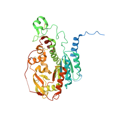
    Asn-linked oligosaccharides are extensively modified during transit through the secretory pathway, first by trimming of the nascent glycan chains and subsequently by initiating and extending multiple oligosaccharide branches from the trimannosyl glycan core. Trimming and branching pathway steps are highly ordered and hierarchal based on the precise substrate specificities of the individual biosynthetic enzymes. A key committed step in the synthesis of complex-type glycans is catalyzed by N -acetylglucosaminyltransferase II (MGAT2), an enzyme that generates the second GlcNAcβ1,2- branch from the trimannosyl glycan core using UDP-GlcNAc as the sugar donor. We determined the structure of human MGAT2 as a Mn 2+ -UDP donor analog complex and as a GlcNAcMan 3 GlcNAc 2 -Asn acceptor complex to reveal the structural basis for substrate recognition and catalysis. The enzyme exhibits a GT-A Rossmann-like fold that employs conserved divalent cation-dependent substrate interactions with the UDP-GlcNAc donor. MGAT2 interactions with the extended glycan acceptor are distinct from other related glycosyltransferases. These interactions are composed of a catalytic subsite that binds the Man-α1,6- monosaccharide acceptor and a distal exosite pocket that binds the GlcNAc-β1,2Man-α1,3Manβ- substrate "recognition arm." Recognition arm interactions are similar to the enzyme-substrate interactions for Golgi α-mannosidase II, a glycoside hydrolase that acts just before MGAT2 in the Asn-linked glycan biosynthetic pathway. These data suggest that substrate binding by MGAT2 employs both conserved and convergent catalytic subsite modules to provide substrate selectivity and catalysis. More broadly, the MGAT2 active-site architecture demonstrates how glycosyltransferases create complementary modular templates for regiospecific extension of glycan structures in mammalian cells.


  • Organizational Affiliation

    Department of Biochemistry and Molecular Biology, University of Georgia, Athens, GA 30602.


Macromolecules
Find similar proteins by:  (by identity cutoff)  |  3D Structure
Entity ID: 1
MoleculeChains Sequence LengthOrganismDetailsImage
Alpha-1,6-mannosyl-glycoprotein 2-beta-N-acetylglucosaminyltransferase
A, B
419Homo sapiensMutation(s): 0 
Gene Names: MGAT2
EC: 2.4.1.143
UniProt & NIH Common Fund Data Resources
Find proteins for Q10469 (Homo sapiens)
Explore Q10469 
Go to UniProtKB:  Q10469
PHAROS:  Q10469
GTEx:  ENSG00000168282 
Entity Groups  
Sequence Clusters30% Identity50% Identity70% Identity90% Identity95% Identity100% Identity
UniProt GroupQ10469
Sequence Annotations
Expand
  • Reference Sequence
Oligosaccharides

Help

Entity ID: 2
MoleculeChains Length2D Diagram Glycosylation3D Interactions
2-acetamido-2-deoxy-beta-D-glucopyranose-(1-2)-alpha-D-mannopyranose-(1-3)-[alpha-D-mannopyranose-(1-6)]beta-D-mannopyranose-(1-4)-2-acetamido-2-deoxy-beta-D-glucopyranose
C, D
5N/A
Glycosylation Resources
GlyTouCan:  G14790EN
GlyCosmos:  G14790EN
GlyGen:  G14790EN
Experimental Data & Validation

Experimental Data

  • Method: X-RAY DIFFRACTION
  • Resolution: 2.80 Å
  • R-Value Free: 0.275 
  • R-Value Work: 0.247 
  • R-Value Observed: 0.249 
  • Space Group: P 2 21 21
Unit Cell:
Length ( Å )Angle ( ˚ )
a = 46.28α = 90
b = 129.16β = 90
c = 175.8γ = 90
Software Package:
Software NamePurpose
PHENIXrefinement
XDSdata reduction
XDSdata scaling
PHENIXphasing

Structure Validation

View Full Validation Report


Ligand Structure Quality Assessment 


Entry History & Funding Information

Deposition Data


Funding OrganizationLocationGrant Number
National Institutes of Health/National Institute of General Medical Sciences (NIH/NIGMS)United States1P01GM107012-02

Revision History  (Full details and data files)

  • Version 1.0: 2018-04-11
    Type: Initial release
  • Version 1.1: 2018-04-18
    Changes: Data collection, Database references
  • Version 1.2: 2018-05-02
    Changes: Data collection, Database references
  • Version 1.3: 2018-05-16
    Changes: Data collection, Database references
  • Version 1.4: 2020-01-01
    Changes: Author supporting evidence
  • Version 2.0: 2020-07-29
    Type: Remediation
    Reason: Carbohydrate remediation
    Changes: Atomic model, Data collection, Derived calculations, Structure summary
  • Version 2.1: 2023-10-04
    Changes: Data collection, Database references, Derived calculations, Refinement description, Structure summary