


Crystal structure of a peptide deformylase from Burkholderia xenovorans in complex with actinonin
Conrady, D.G., Abendroth, J., Lorimer, D.D., Edwards, T.E.To be published.
Experimental Data Snapshot
Starting Model: experimental
View more details
Entity ID: 1 | |||||
|---|---|---|---|---|---|
| Molecule | Chains | Sequence Length | Organism | Details | Image |
| Peptide deformylase | 185 | Paraburkholderia xenovorans LB400 | Mutation(s): 0 Gene Names: def, Bxe_A1677 EC: 3.5.1.88 | 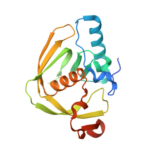 | |
UniProt | |||||
Entity Groups | |||||
| Sequence Clusters | 30% Identity50% Identity70% Identity90% Identity95% Identity100% Identity | ||||
| UniProt Group | Q13XB1 | ||||
Sequence AnnotationsExpand | |||||
Reference Sequence | |||||
| Ligands 3 Unique | |||||
|---|---|---|---|---|---|
| ID | Chains | Name / Formula / InChI Key | 2D Diagram | 3D Interactions | |
| BB2 Download:Ideal Coordinates CCD File | F [auth A], I [auth B], M [auth C], O [auth D] | ACTINONIN C19 H35 N3 O5 XJLATMLVMSFZBN-VYDXJSESSA-N |  | ||
| FE2 Download:Ideal Coordinates CCD File | E [auth A], H [auth B], L [auth C], N [auth D] | FE (II) ION Fe CWYNVVGOOAEACU-UHFFFAOYSA-N |  | ||
| MG Download:Ideal Coordinates CCD File | G [auth A], J [auth B], K [auth B] | MAGNESIUM ION Mg JLVVSXFLKOJNIY-UHFFFAOYSA-N |  | ||
| Length ( Å ) | Angle ( ˚ ) |
|---|---|
| a = 63.11 | α = 90 |
| b = 90.33 | β = 90 |
| c = 140.21 | γ = 90 |
| Software Name | Purpose |
|---|---|
| XSCALE | data scaling |
| PHASER | phasing |
| PHENIX | refinement |
| PDB_EXTRACT | data extraction |
| XDS | data reduction |
| PHASER | phasing |


