5VAW | pdb_00005vaw

Fusion of Maltose-binding Protein and PilA from Acinetobacter baumannii AB5075


Experimental Data Snapshot

  • Method: X-RAY DIFFRACTION
  • Resolution: 1.69 Å
  • R-Value Free: 
    0.242 (Depositor), 0.242 (DCC) 
  • R-Value Work: 
    0.209 (Depositor), 0.209 (DCC) 
  • R-Value Observed: 
    0.210 (Depositor) 

Starting Model: experimental
View more details

wwPDB Validation 3D Report Full Report

Validation slider image for 5VAW

This is version 1.4 of the entry. See complete history

Literature

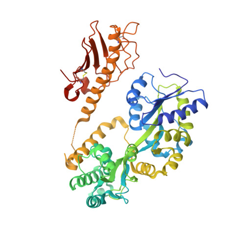
The structure of PilA fromAcinetobacter baumanniiAB5075 suggests a mechanism for functional specialization inAcinetobactertype IV pili.

Ronish, L.A.Lillehoj, E.Fields, J.K.Sundberg, E.J.Piepenbrink, K.H.

(2019) J Biological Chem 294: 218-230

  • DOI: https://doi.org/10.1074/jbc.RA118.005814
  • Primary Citation Related Structures: 
    5VAW

  • PubMed Abstract: 

    Type IV pili (T4P) are bacterial appendages composed of protein subunits, called pilins, noncovalently assembled into helical fibers. T4P are essential, in many bacterial species, for processes as diverse as twitching motility, natural competence, biofilm or microcolony formation, and host cell adhesion. The genes encoding type IV pili are found universally in the Gram-negative, aerobic, nonflagellated, and pathogenic coccobacillus Acinetobacter baumannii , but there is considerable variation in PilA, the major protein subunit, both in amino acid sequence and in glycosylation patterns. Here we report the X-ray crystal structure of PilA from AB5075, a recently characterized, highly virulent isolate, at 1.9 Å resolution and compare it to homologues from A. baumannii strains ACICU and BIDMC57, which are C-terminally glycosylated. These structural comparisons revealed that PilA AB5075 exhibits a distinctly electronegative surface chemistry. To understand the functional consequences of this change in surface electrostatics, we complemented a Δ pilA knockout strain with divergent pilA genes from ACICU, BIDMC57, and AB5075. The resulting transgenic strains showed differential twitching motility and biofilm formation while maintaining the ability to adhere to epithelial cells. PilA AB5075 and PilA ACICU , although structurally similar, promote different characteristics, favoring twitching motility and biofilm formation, respectively. These results support a model in which differences in pilus electrostatics affect the equilibrium of microcolony formation, which in turn alters the balance between motility and biofilm formation in Acinetobacter .


  • Organizational Affiliation
    • Department of Biochemistry, University of Nebraska-Lincoln, Lincoln, Nebraska 68588.

Macromolecule Content 

  • Total Structure Weight: 54.68 kDa 
  • Atom Count: 3,966 
  • Modeled Residue Count: 484 
  • Deposited Residue Count: 508 
  • Unique protein chains: 1

Macromolecules

Find similar proteins by:|  3D Structure
Entity ID: 1
MoleculeChains  Sequence LengthOrganismDetailsImage
Maltose-binding periplasmic protein,Type IV pilin PilA508Escherichia coliAcinetobacter baumannii
This entity is chimeric
Mutation(s): 11 
UniProt
Find proteins for A0A0D5YCX7 (Acinetobacter baumannii)
Explore A0A0D5YCX7 
Go to UniProtKB:  A0A0D5YCX7
Find proteins for P0AEX9 (Escherichia coli (strain K12))
Explore P0AEX9 
Go to UniProtKB:  P0AEX9
Entity Groups
Sequence Clusters30% Identity50% Identity70% Identity90% Identity95% Identity100% Identity
UniProt GroupsA0A0D5YCX7P0AEX9
Sequence Annotations
Expand
Reference Sequence

Small Molecules

Ligands 5 Unique
IDChains Name / Formula / InChI Key2D Diagram3D Interactions
LYS

Query on LYS


Download:Ideal Coordinates CCD File
E [auth A]LYSINE
C6 H15 N2 O2
KDXKERNSBIXSRK-YFKPBYRVSA-O
GLU

Query on GLU


Download:Ideal Coordinates CCD File
D [auth A]GLUTAMIC ACID
C5 H9 N O4
WHUUTDBJXJRKMK-VKHMYHEASA-N
MPD

Query on MPD


Download:Ideal Coordinates CCD File
B [auth A](4S)-2-METHYL-2,4-PENTANEDIOL
C6 H14 O2
SVTBMSDMJJWYQN-YFKPBYRVSA-N
SER

Query on SER


Download:Ideal Coordinates CCD File
F [auth A]SERINE
C3 H7 N O3
MTCFGRXMJLQNBG-REOHCLBHSA-N
NA

Query on NA


Download:Ideal Coordinates CCD File
C [auth A]SODIUM ION
Na
FKNQFGJONOIPTF-UHFFFAOYSA-N

Experimental Data & Validation

Experimental Data

  • Method: X-RAY DIFFRACTION
  • Resolution: 1.69 Å
  • R-Value Free:  0.242 (Depositor), 0.242 (DCC) 
  • R-Value Work:  0.209 (Depositor), 0.209 (DCC) 
  • R-Value Observed: 0.210 (Depositor) 
Space Group: P 1 21 1
Unit Cell:
Length ( Å )Angle ( ˚ )
a = 39.557α = 90
b = 103.04β = 98.95
c = 56.195γ = 90
Software Package:
Software NamePurpose
PHENIXrefinement
Aimlessdata scaling
PHASERphasing
PDB_EXTRACTdata extraction
XDSdata reduction

Structure Validation

View Full Validation Report


Entry History 

Deposition Data

Revision History  (Full details and data files)

  • Version 1.0: 2018-03-28
    Type: Initial release
  • Version 1.1: 2018-11-28
    Changes: Data collection, Database references
  • Version 1.2: 2019-01-16
    Changes: Data collection, Database references
  • Version 1.3: 2023-10-04
    Changes: Data collection, Database references, Derived calculations, Refinement description
  • Version 1.4: 2024-11-13
    Changes: Structure summary