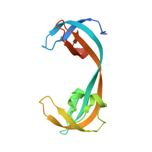
5VAR

Crystal structure of KDM4A tandem TUDOR domain in complex with a tri-methyl lysine competitive inhibitor


Experimental Data Snapshot

  • Method: X-RAY DIFFRACTION
  • Resolution: 1.83 Å
  • R-Value Free: 0.243 
  • R-Value Work: 0.194 
  • R-Value Observed: 0.196 

wwPDB Validation   3D Report Full Report


This is version 1.4 of the entry. See complete history


Literature

Targeting lysine specific demethylase 4A (KDM4A) tandem TUDOR domain - A fragment based approach.

Upadhyay, A.K.Judge, R.A.Li, L.Pithawalla, R.Simanis, J.Bodelle, P.M.Marin, V.L.Henry, R.F.Petros, A.M.Sun, C.

(2018) Bioorg Med Chem Lett 28: 1708-1713

  • DOI: https://doi.org/10.1016/j.bmcl.2018.04.050
  • Primary Citation of Related Structures:  
    5VAR

  • PubMed Abstract: 

    The tandem TUDOR domains present in the non-catalytic C-terminal half of the KDM4A, 4B and 4C enzymes play important roles in regulating their chromatin localizations and substrate specificities. They achieve this regulatory role by binding to different tri-methylated lysine residues on histone H3 (H3-K4me3, H3-K23me3) and histone H4 (H4-K20me3) depending upon the specific chromatin environment. In this work, we have used a 2D-NMR based fragment screening approach to identify a novel fragment (1a), which binds to the KDM4A-TUDOR domain and shows modest competition with H3-K4me3 binding in biochemical as well as in vitro cell based assays. A co-crystal structure of KDM4A TUDOR domain in complex with 1a shows that the fragment binds stereo-specifically to the methyl lysine binding pocket forming a network of strong hydrogen bonds and hydrophobic interactions. We anticipate that the fragment 1a can be further developed into a novel allosteric inhibitor of the KDM4 family of enzymes through targeting their C-terminal tandem TUDOR domain.


  • Organizational Affiliation

    AbbVie Inc., 1 North Waukegan Road, North Chicago, IL 60064, USA. Electronic address: anup.upadhyay@abbvie.com.


Macromolecules
Find similar proteins by:  (by identity cutoff)  |  3D Structure
Entity ID: 1
MoleculeChains Sequence LengthOrganismDetailsImage
Lysine-specific demethylase 4A119Homo sapiensMutation(s): 0 
Gene Names: KDM4AJHDM3AJMJD2JMJD2AKIAA0677
EC: 1.14.11 (PDB Primary Data), 1.14.11.66 (UniProt), 1.14.11.69 (UniProt)
UniProt & NIH Common Fund Data Resources
Find proteins for O75164 (Homo sapiens)
Explore O75164 
Go to UniProtKB:  O75164
PHAROS:  O75164
Entity Groups  
Sequence Clusters30% Identity50% Identity70% Identity90% Identity95% Identity100% Identity
UniProt GroupO75164
Sequence Annotations
Expand
  • Reference Sequence
Small Molecules
Ligands 1 Unique
IDChains Name / Formula / InChI Key2D Diagram3D Interactions
92Y
Query on 92Y

Download Ideal Coordinates CCD File 
B [auth A](1R,2S,3R,4S)-3-[(dimethylamino)methyl]-1-phenylbicyclo[2.2.1]heptan-2-ol
C16 H23 N O
HGNFUCJMHJYHIN-QCEMKRCNSA-N
Binding Affinity Annotations 
IDSourceBinding Affinity
92Y BindingDB:  5VAR Kd: 8.00e+4 (nM) from 1 assay(s)
Binding MOAD:  5VAR Kd: 8.17e+4 (nM) from 1 assay(s)
Experimental Data & Validation

Experimental Data

  • Method: X-RAY DIFFRACTION
  • Resolution: 1.83 Å
  • R-Value Free: 0.243 
  • R-Value Work: 0.194 
  • R-Value Observed: 0.196 
  • Space Group: P 1 21 1
Unit Cell:
Length ( Å )Angle ( ˚ )
a = 33.27α = 90
b = 39.84β = 101.61
c = 44.3γ = 90
Software Package:
Software NamePurpose
Aimlessdata scaling
MOLREPphasing
BUSTERrefinement
PDB_EXTRACTdata extraction
XDSdata reduction

Structure Validation

View Full Validation Report


Entry History 

Deposition Data

Revision History  (Full details and data files)

  • Version 1.0: 2018-03-28
    Type: Initial release
  • Version 1.1: 2018-05-09
    Changes: Data collection, Database references
  • Version 1.2: 2018-05-16
    Changes: Data collection, Database references
  • Version 1.3: 2018-09-05
    Changes: Data collection, Database references
  • Version 1.4: 2023-10-04
    Changes: Data collection, Database references, Refinement description