5VAC

Crystal Structure of ATXR5 SET domain in complex with K36me3 histone H3 peptide


Experimental Data Snapshot

  • Method: X-RAY DIFFRACTION
  • Resolution: 1.95 Å
  • R-Value Free: 0.228 
  • R-Value Work: 0.189 
  • R-Value Observed: 0.191 

wwPDB Validation   3D Report Full Report


Ligand Structure Quality Assessment 


This is version 1.2 of the entry. See complete history


Literature

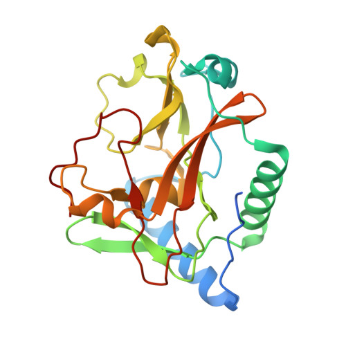
Molecular basis for the methylation specificity of ATXR5 for histone H3.

Bergamin, E.Sarvan, S.Malette, J.Eram, M.S.Yeung, S.Mongeon, V.Joshi, M.Brunzelle, J.S.Michaels, S.D.Blais, A.Vedadi, M.Couture, J.F.

(2017) Nucleic Acids Res 45: 6375-6387

  • DOI: https://doi.org/10.1093/nar/gkx224
  • Primary Citation of Related Structures:  
    5VA6, 5VAB, 5VAC, 5VAH, 5VBC

  • PubMed Abstract: 

    In plants, the histone H3.1 lysine 27 (H3K27) mono-methyltransferases ARABIDOPSIS TRITHORAX RELATED PROTEIN 5 and 6 (ATXR5/6) regulate heterochromatic DNA replication and genome stability. Our initial studies showed that ATXR5/6 discriminate between histone H3 variants and preferentially methylate K27 on H3.1. In this study, we report three regulatory mechanisms contributing to the specificity of ATXR5/6. First, we show that ATXR5 preferentially methylates the R/F-K*-S/C-G/A-P/C motif with striking preference for hydrophobic and aromatic residues in positions flanking this core of five amino acids. Second, we demonstrate that post-transcriptional modifications of residues neighboring K27 that are typically associated with actively transcribed chromatin are detrimental to ATXR5 activity. Third, we show that ATXR5 PHD domain employs a narrow binding pocket to selectively recognize unmethylated K4 of histone H3. Finally, we demonstrate that deletion or mutation of the PHD domain reduces the catalytic efficiency (kcat/Km of AdoMet) of ATXR5 up to 58-fold, highlighting the multifunctional nature of ATXR5 PHD domain. Overall, our results suggest that several molecular determinants regulate ATXR5/6 methyltransferase activity and epigenetic inheritance of H3.1 K27me1 mark in plants.


  • Organizational Affiliation

    Ottawa Institute of Systems Biology, Department of Biochemistry, Microbiology and Immunology, University of Ottawa, 451 Smyth Road, Ottawa, Ontario K1H 8M5, Canada.


Macromolecules
Find similar proteins by:  (by identity cutoff)  |  3D Structure
Entity ID: 1
MoleculeChains Sequence LengthOrganismDetailsImage
Probable Histone-lysine N-methyltransferase ATXR5229Ricinus communisMutation(s): 0 
Gene Names: ATXR5RCOM_1460410
EC: 2.1.1.43
UniProt
Find proteins for B9RU15 (Ricinus communis)
Explore B9RU15 
Go to UniProtKB:  B9RU15
Entity Groups  
Sequence Clusters30% Identity50% Identity70% Identity90% Identity95% Identity100% Identity
UniProt GroupB9RU15
Sequence Annotations
Expand
  • Reference Sequence

Find similar proteins by:  Sequence   |   3D Structure  

Entity ID: 2
MoleculeChains Sequence LengthOrganismDetailsImage
Histone H3.2B [auth C]19Homo sapiensMutation(s): 0 
UniProt & NIH Common Fund Data Resources
Find proteins for Q71DI3 (Homo sapiens)
Explore Q71DI3 
Go to UniProtKB:  Q71DI3
PHAROS:  Q71DI3
Entity Groups  
Sequence Clusters30% Identity50% Identity70% Identity90% Identity95% Identity100% Identity
UniProt GroupQ71DI3
Sequence Annotations
Expand
  • Reference Sequence
Small Molecules
Ligands 2 Unique
IDChains Name / Formula / InChI Key2D Diagram3D Interactions
SAH
Query on SAH

Download Ideal Coordinates CCD File 
C [auth A]S-ADENOSYL-L-HOMOCYSTEINE
C14 H20 N6 O5 S
ZJUKTBDSGOFHSH-WFMPWKQPSA-N
DMS
Query on DMS

Download Ideal Coordinates CCD File 
D [auth A]DIMETHYL SULFOXIDE
C2 H6 O S
IAZDPXIOMUYVGZ-UHFFFAOYSA-N
Experimental Data & Validation

Experimental Data

  • Method: X-RAY DIFFRACTION
  • Resolution: 1.95 Å
  • R-Value Free: 0.228 
  • R-Value Work: 0.189 
  • R-Value Observed: 0.191 
  • Space Group: I 2 2 2
Unit Cell:
Length ( Å )Angle ( ˚ )
a = 74.548α = 90
b = 81.353β = 90
c = 88.503γ = 90
Software Package:
Software NamePurpose
PHENIXrefinement
XDSdata reduction
Aimlessdata scaling
PHASERphasing

Structure Validation

View Full Validation Report


Ligand Structure Quality Assessment 


Entry History 

Deposition Data

Revision History  (Full details and data files)

  • Version 1.0: 2017-04-19
    Type: Initial release
  • Version 1.1: 2017-09-20
    Changes: Database references
  • Version 1.2: 2023-10-04
    Changes: Data collection, Database references, Refinement description