5V6I

Glycan binding protein Y3 from mushroom Coprinus comatus possesses anti-leukemic activity - Pt derivative


Experimental Data Snapshot

  • Method: X-RAY DIFFRACTION
  • Resolution: 1.70 Å
  • R-Value Free: 0.194 
  • R-Value Work: 0.160 
  • R-Value Observed: 0.161 

wwPDB Validation   3D Report Full Report


This is version 2.0 of the entry. See complete history


Literature

Cytotoxic protein from the mushroom Coprinus comatus possesses a unique mode for glycan binding and specificity.

Zhang, P.Li, K.Yang, G.Xia, C.Polston, J.E.Li, G.Li, S.Lin, Z.Yang, L.J.Bruner, S.D.Ding, Y.

(2017) Proc Natl Acad Sci U S A 114: 8980-8985

  • DOI: https://doi.org/10.1073/pnas.1706894114
  • Primary Citation of Related Structures:  
    5V6I, 5V6J

  • PubMed Abstract: 

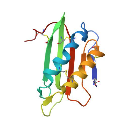
    Glycans possess significant chemical diversity; glycan binding proteins (GBPs) recognize specific glycans to translate their structures to functions in various physiological and pathological processes. Therefore, the discovery and characterization of novel GBPs and characterization of glycan-GBP interactions are significant to provide potential targets for therapeutic intervention of many diseases. Here, we report the biochemical, functional, and structural characterization of a 130-amino-acid protein, Y3, from the mushroom Coprinus comatus Biochemical studies of recombinant Y3 from a yeast expression system demonstrated the protein is a unique GBP. Additionally, we show that Y3 exhibits selective and potent cytotoxicity toward human T-cell leukemia Jurkat cells compared with a panel of cancer cell lines via inducing caspase-dependent apoptosis. Screening of a glycan array demonstrated GalNAcβ1-4(Fucα1-3)GlcNAc (LDNF) as a specific Y3-binding ligand. To provide a structural basis for function, the crystal structure was solved to a resolution of 1.2 Å, revealing a single-domain αβα-sandwich motif. Two monomers were dimerized to form a large 10-stranded, antiparallel β-sheet flanked by α-helices on each side, representing a unique oligomerization mode among GBPs. A large glycan binding pocket extends into the dimeric interface, and docking of LDNF identified key residues for glycan interactions. Disruption of residues predicted to be involved in LDNF/Y3 interactions resulted in the significant loss of binding to Jurkat T-cells and severely impaired their cytotoxicity. Collectively, these results demonstrate Y3 to be a GBP with selective cytotoxicity toward human T-cell leukemia cells and indicate its potential use in cancer diagnosis and treatment.


  • Organizational Affiliation

    Department of Medicinal Chemistry, Center for Natural Products, Drug Discovery and Development, University of Florida, Gainesville, FL 32610.


Macromolecules
Find similar proteins by:  (by identity cutoff)  |  3D Structure
Entity ID: 1
MoleculeChains Sequence LengthOrganismDetailsImage
TMV resistance protein Y3
A, B
112Coprinus comatusMutation(s): 0 
Gene Names: y3
UniProt
Find proteins for G3BK00 (Coprinus comatus)
Explore G3BK00 
Go to UniProtKB:  G3BK00
Entity Groups  
Sequence Clusters30% Identity50% Identity70% Identity90% Identity95% Identity100% Identity
UniProt GroupG3BK00
Sequence Annotations
Expand
  • Reference Sequence
Small Molecules
Ligands 3 Unique
IDChains Name / Formula / InChI Key2D Diagram3D Interactions
NHE
Query on NHE

Download Ideal Coordinates CCD File 
C [auth A],
T [auth B]
2-[N-CYCLOHEXYLAMINO]ETHANE SULFONIC ACID
C8 H17 N O3 S
MKWKNSIESPFAQN-UHFFFAOYSA-N
PT
Query on PT

Download Ideal Coordinates CCD File 
D [auth B],
E [auth B],
F [auth B],
G [auth B]
PLATINUM (II) ION
Pt
HRGDZIGMBDGFTC-UHFFFAOYSA-N
CL
Query on CL

Download Ideal Coordinates CCD File 
H [auth B]
I [auth B]
J [auth B]
K [auth B]
L [auth B]
H [auth B],
I [auth B],
J [auth B],
K [auth B],
L [auth B],
M [auth B],
N [auth B],
O [auth B],
P [auth B],
Q [auth B],
R [auth B],
S [auth B]
CHLORIDE ION
Cl
VEXZGXHMUGYJMC-UHFFFAOYSA-M
Modified Residues  1 Unique
IDChains TypeFormula2D DiagramParent
PCA
Query on PCA
A, B
L-PEPTIDE LINKINGC5 H7 N O3GLN
Experimental Data & Validation

Experimental Data

  • Method: X-RAY DIFFRACTION
  • Resolution: 1.70 Å
  • R-Value Free: 0.194 
  • R-Value Work: 0.160 
  • R-Value Observed: 0.161 
  • Space Group: P 1 21 1
Unit Cell:
Length ( Å )Angle ( ˚ )
a = 41.102α = 90
b = 55.237β = 99.52
c = 41.879γ = 90
Software Package:
Software NamePurpose
XDSdata reduction
Aimlessdata scaling
SHELXDEphasing
PHENIXrefinement

Structure Validation

View Full Validation Report


Entry History & Funding Information

Deposition Data


Funding OrganizationLocationGrant Number
National Institutes of Health/National Institute of General Medical Sciences (NIH/NIGMS)United StatesR24 GM098791
University of FloridaUnited States--

Revision History  (Full details and data files)

  • Version 1.0: 2017-08-16
    Type: Initial release
  • Version 1.1: 2017-08-23
    Changes: Author supporting evidence, Database references
  • Version 1.2: 2017-09-06
    Changes: Database references
  • Version 2.0: 2020-01-01
    Changes: Author supporting evidence, Polymer sequence