Structural basis of mammalian glycan targeting by Vibrio cholerae cytolysin and biofilm proteins.
De, S., Kaus, K., Sinclair, S., Case, B.C., Olson, R.(2018) PLoS Pathog 14: e1006841-e1006841
- PubMed: 29432487 Search on PubMedSearch on PubMed Central
- DOI: https://doi.org/10.1371/journal.ppat.1006841
- Primary Citation Related Structures:
5V6C, 5V6F, 5V6K - PubMed Abstract:
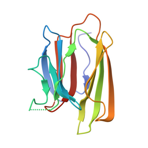
Vibrio cholerae is an aquatic gram-negative microbe responsible for cholera, a pandemic disease causing life-threatening diarrheal outbreaks in populations with limited access to health care. Like most pathogenic bacteria, V. cholerae secretes virulence factors to assist colonization of human hosts, several of which bind carbohydrate receptors found on cell-surfaces. Understanding how pathogenic virulence proteins specifically target host cells is important for the development of treatment strategies to fight bacterial infections. Vibrio cholerae cytolysin (VCC) is a secreted pore-forming toxin with a carboxy-terminal β-prism domain that targets complex N-glycans found on mammalian cell-surface proteins. To investigate glycan selectivity, we studied the VCC β-prism domain and two additional β-prism domains found within the V. cholerae biofilm matrix protein RbmC. We show that the two RbmC β-prism domains target a similar repertoire of complex N-glycan receptors as VCC and find through binding and modeling studies that a branched pentasaccharide core (GlcNAc2-Man3) represents the likely footprint interacting with these domains. To understand the structural basis of V. cholerae β-prism selectivity, we solved high-resolution crystal structures of fragments of the pentasaccharide core bound to one RbmC β-prism domain and conducted mutagenesis experiments on the VCC toxin. Our results highlight a common strategy for cell-targeting utilized by both toxin and biofilm matrix proteins in Vibrio cholerae and provide a structural framework for understanding the specificity for individual receptors. Our results suggest that a common strategy for disrupting carbohydrate interactions could affect multiple virulence factors produced by V. cholerae, as well as similar β-prism domains found in other vibrio pathogens.
- Department of Molecular Biology and Biochemistry, Molecular Biophysics Program, Wesleyan University, Middletown, Connecticut, United States of America.
Organizational Affiliation: