5V52

Structure of TIGIT bound to nectin-2 (CD112)


Experimental Data Snapshot

  • Method: X-RAY DIFFRACTION
  • Resolution: 3.10 Å
  • R-Value Free: 0.236 
  • R-Value Work: 0.218 
  • R-Value Observed: 0.218 

wwPDB Validation   3D Report Full Report


Ligand Structure Quality Assessment 


This is version 1.7 of the entry. See complete history


Literature

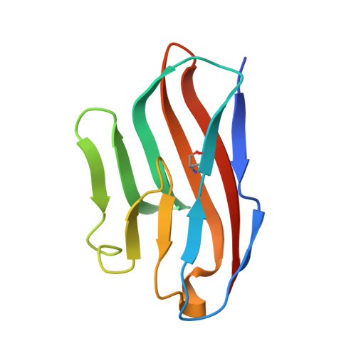
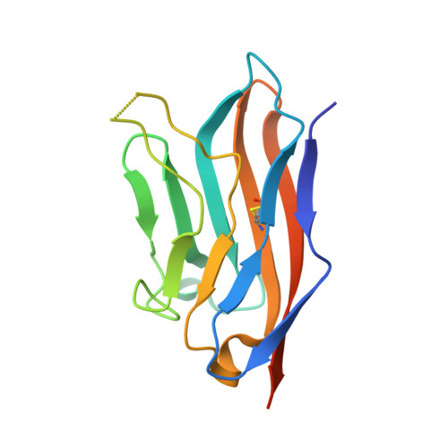
Recognition of nectin-2 by the natural killer cell receptor T cell immunoglobulin and ITIM domain (TIGIT).

Deuss, F.A.Gully, B.S.Rossjohn, J.Berry, R.

(2017) J Biol Chem 292: 11413-11422

  • DOI: https://doi.org/10.1074/jbc.M117.786483
  • Primary Citation of Related Structures:  
    5V52

  • PubMed Abstract: 
  • T cell immunoglobulin and ITIM domain (TIGIT) is an inhibitory receptor expressed on the surface of natural killer (NK) cells. TIGIT recognizes nectin and nectin-like adhesion molecules and thus plays a critical role in the innate immune response to malignant transformation ...

    T cell immunoglobulin and ITIM domain (TIGIT) is an inhibitory receptor expressed on the surface of natural killer (NK) cells. TIGIT recognizes nectin and nectin-like adhesion molecules and thus plays a critical role in the innate immune response to malignant transformation. Although the TIGIT nectin-like protein-5 (necl-5) interaction is well understood, how TIGIT engages nectin-2, a receptor that is broadly over-expressed in breast and ovarian cancer, remains unknown. Here, we show that TIGIT bound to the immunoglobulin domain of nectin-2 that is most distal from the membrane with an affinity of 6 μm, which was moderately lower than the affinity observed for the TIGIT/necl-5 interaction (3.2 μm). The TIGIT/nectin-2 binding disrupted pre-assembled nectin-2 oligomers, suggesting that receptor-ligand and ligand-ligand associations are mutually exclusive events. Indeed, the crystal structure of TIGIT bound to the first immunoglobulin domain of nectin-2 indicated that the receptor and ligand dock using the same molecular surface and a conserved "lock and key" binding motifs previously observed to mediate nectin/nectin homotypic interactions as well as TIGIT/necl-5 recognition. Using a mutagenesis approach, we dissected the energetic basis for the TIGIT/nectin-2 interaction and revealed that an "aromatic key" of nectin-2 is critical for this interaction, whereas variations in the lock were tolerated. Moreover, we found that the C-C' loop of the ligand dictates the TIGIT binding hierarchy. Altogether, these findings broaden our understanding of nectin/nectin receptor interactions and have implications for better understanding the molecular basis for autoimmune disease and cancer.


    Organizational Affiliation

    the Australian Research Council Centre of Excellence in Advanced Molecular Imaging, Monash University, Clayton, Victoria 3800, Australia, and.


Macromolecules
Find similar proteins by:  (by identity cutoff)  |  3D Structure
Entity ID: 1
MoleculeChainsSequence LengthOrganismDetailsImage
T-cell immunoreceptor with Ig and ITIM domains
A, B
107Homo sapiensMutation(s): 1 
Gene Names: TIGITVSIG9VSTM3
UniProt & NIH Common Fund Data Resources
Find proteins for Q495A1 (Homo sapiens)
Explore Q495A1 
Go to UniProtKB:  Q495A1
PHAROS:  Q495A1
Entity Groups  
Sequence Clusters30% Identity50% Identity70% Identity90% Identity95% Identity100% Identity
UniProt GroupQ495A1
Protein Feature View
Expand
  • Reference Sequence
Find similar proteins by:  (by identity cutoff)  |  3D Structure
Entity ID: 2
MoleculeChainsSequence LengthOrganismDetailsImage
Nectin-2
C, D
140Homo sapiensMutation(s): 0 
Gene Names: NECTIN2HVEBPRR2PVRL2
UniProt & NIH Common Fund Data Resources
Find proteins for Q92692 (Homo sapiens)
Explore Q92692 
Go to UniProtKB:  Q92692
PHAROS:  Q92692
Entity Groups  
Sequence Clusters30% Identity50% Identity70% Identity90% Identity95% Identity100% Identity
UniProt GroupQ92692
Protein Feature View
Expand
  • Reference Sequence
Small Molecules
Ligands 3 Unique
IDChainsName / Formula / InChI Key2D Diagram3D Interactions
NAG
Query on NAG

Download Ideal Coordinates CCD File 
G [auth C]2-acetamido-2-deoxy-beta-D-glucopyranose
C8 H15 N O6
OVRNDRQMDRJTHS-FMDGEEDCSA-N
 Ligand Interaction
SO4
Query on SO4

Download Ideal Coordinates CCD File 
F [auth B]SULFATE ION
O4 S
QAOWNCQODCNURD-UHFFFAOYSA-L
 Ligand Interaction
GOL
Query on GOL

Download Ideal Coordinates CCD File 
E [auth A]GLYCEROL
C3 H8 O3
PEDCQBHIVMGVHV-UHFFFAOYSA-N
 Ligand Interaction
Experimental Data & Validation

Experimental Data

  • Method: X-RAY DIFFRACTION
  • Resolution: 3.10 Å
  • R-Value Free: 0.236 
  • R-Value Work: 0.218 
  • R-Value Observed: 0.218 
  • Space Group: P 43 21 2
Unit Cell:
Length ( Å )Angle ( ˚ )
a = 68.5α = 90
b = 68.5β = 90
c = 253.91γ = 90
Software Package:
Software NamePurpose
BUSTERrefinement
iMOSFLMdata reduction
SCALAdata scaling
PHASERphasing

Structure Validation

View Full Validation Report


Ligand Structure Quality Assessment 


Entry History & Funding Information

Deposition Data


Funding OrganizationLocationGrant Number
National Health and Medical Research Council (NHMRC, Australia)AustraliaAPP1109901

Revision History  (Full details and data files)

  • Version 1.0: 2017-05-24
    Type: Initial release
  • Version 1.1: 2017-06-07
    Changes: Database references
  • Version 1.2: 2017-07-19
    Changes: Database references
  • Version 1.3: 2017-09-20
    Changes: Author supporting evidence
  • Version 1.4: 2020-01-08
    Changes: Author supporting evidence
  • Version 1.5: 2020-02-26
    Changes: Data collection
  • Version 1.6: 2020-07-29
    Type: Remediation
    Reason: Carbohydrate remediation
    Changes: Data collection, Derived calculations, Structure summary
  • Version 1.7: 2023-10-04
    Changes: Data collection, Database references, Refinement description, Structure summary