5V4K

Crystal structure of NEDD4 LIR-fused human LC3B_2-119


Experimental Data Snapshot

  • Method: X-RAY DIFFRACTION
  • Resolution: 2.10 Å
  • R-Value Free: 0.233 
  • R-Value Work: 0.182 
  • R-Value Observed: 0.184 

wwPDB Validation   3D Report Full Report


This is version 1.2 of the entry. See complete history


Literature

Insights into links between autophagy and the ubiquitin system from the structure of LC3B bound to the LIR motif from the E3 ligase NEDD4.

Qiu, Y.Zheng, Y.Wu, K.P.Schulman, B.A.

(2017) Protein Sci 26: 1674-1680

  • DOI: https://doi.org/10.1002/pro.3186
  • Primary Citation of Related Structures:  
    5V4K

  • PubMed Abstract: 

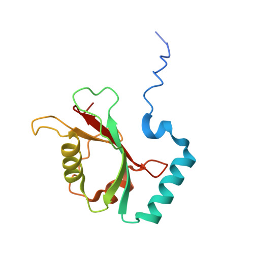
    Members of the LC3/GABARAP family of ubiquitin-like proteins function during autophagy by serving as membrane linked protein-binding platforms. Their C-termini are physically attached to membranes through covalent linkage to primary amines on lipids such as phosphatidylethanolamine, while their ubiquitin-like fold domains bind "LIR" (LC3-Interacting Region) sequences found within an extraordinarily diverse array of proteins including regulators of autophagy, adaptors that recruit ubiquitinated cargoes to be degraded, and even proteins controlling processes at membranes that are not associated with autophagy. Recently, LC3/GABARAP proteins were found to bind the ubiquitin E3 ligase NEDD4 to influence ubiquitination associated with autophagy in human cell lines. Here, we use purified recombinant proteins to define LC3B interactions with a specific LIR sequence from NEDD4, present a crystal structure showing atomic details of the interaction, and show that LC3B-binding can steer intrinsic NEDD4 E3 ligase activity. The data provide detailed molecular insights underlying recruitment of an E3 ubiquitin ligase to phagophores during autophagy.


  • Organizational Affiliation

    Department of Structural Biology, St. Jude Children's Research Hospital, Memphis, Tennessee, 38105.


Macromolecules
Find similar proteins by:  (by identity cutoff)  |  3D Structure
Entity ID: 1
MoleculeChains Sequence LengthOrganismDetailsImage
Microtubule-associated proteins 1A/1B light chain 3B,Microtubule-associated proteins 1A/1B light chain 3B,Microtubule-associated proteins 1A/1B light chain 3B
A, B
132Homo sapiensMutation(s): 0 
Gene Names: MAP1LC3BMAP1ALC3
UniProt & NIH Common Fund Data Resources
Find proteins for Q9GZQ8 (Homo sapiens)
Explore Q9GZQ8 
Go to UniProtKB:  Q9GZQ8
PHAROS:  Q9GZQ8
GTEx:  ENSG00000140941 
Entity Groups  
Sequence Clusters30% Identity50% Identity70% Identity90% Identity95% Identity100% Identity
UniProt GroupQ9GZQ8
Sequence Annotations
Expand
  • Reference Sequence
Experimental Data & Validation

Experimental Data

  • Method: X-RAY DIFFRACTION
  • Resolution: 2.10 Å
  • R-Value Free: 0.233 
  • R-Value Work: 0.182 
  • R-Value Observed: 0.184 
  • Space Group: P 43 21 2
Unit Cell:
Length ( Å )Angle ( ˚ )
a = 69.57α = 90
b = 69.57β = 90
c = 119.62γ = 90
Software Package:
Software NamePurpose
PHENIXrefinement
XDSdata reduction
Aimlessdata scaling
PHASERphasing

Structure Validation

View Full Validation Report


Entry History & Funding Information

Deposition Data


Funding OrganizationLocationGrant Number
National Institutes of Health/National Institute of General Medical Sciences (NIH/NIGMS)United States--

Revision History  (Full details and data files)

  • Version 1.0: 2017-05-17
    Type: Initial release
  • Version 1.1: 2017-08-02
    Changes: Author supporting evidence, Database references
  • Version 1.2: 2022-04-13
    Changes: Author supporting evidence, Database references