5V43 | pdb_00005v43

Engineered human IgG Fc domain aglyco801


Experimental Data Snapshot

  • Method: X-RAY DIFFRACTION
  • Resolution: 2.32 Å
  • R-Value Free: 
    0.225 (Depositor), 0.248 (DCC) 
  • R-Value Work: 
    0.195 (Depositor), 0.214 (DCC) 
  • R-Value Observed: 
    0.197 (Depositor) 

wwPDB Validation 3D Report Full Report

Validation slider image for 5V43

This is version 1.4 of the entry. See complete history

Literature

IgG Fc domains that bind C1q but not effector Fc gamma receptors delineate the importance of complement-mediated effector functions.

Lee, C.H.Romain, G.Yan, W.Watanabe, M.Charab, W.Todorova, B.Lee, J.Triplett, K.Donkor, M.Lungu, O.I.Lux, A.Marshall, N.Lindorfer, M.A.Goff, O.R.Balbino, B.Kang, T.H.Tanno, H.Delidakis, G.Alford, C.Taylor, R.P.Nimmerjahn, F.Varadarajan, N.Bruhns, P.Zhang, Y.J.Georgiou, G.

(2017) Nat Immunol 18: 889-898

  • DOI: https://doi.org/10.1038/ni.3770
  • Primary Citation Related Structures: 
    5V43, 5V4E

  • PubMed Abstract: 

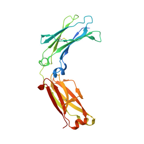
    Engineered crystallizable fragment (Fc) regions of antibody domains, which assume a unique and unprecedented asymmetric structure within the homodimeric Fc polypeptide, enable completely selective binding to the complement component C1q and activation of complement via the classical pathway without any concomitant engagement of the Fcγ receptor (FcγR). We used the engineered Fc domains to demonstrate in vitro and in mouse models that for therapeutic antibodies, complement-dependent cell-mediated cytotoxicity (CDCC) and complement-dependent cell-mediated phagocytosis (CDCP) by immunological effector molecules mediated the clearance of target cells with kinetics and efficacy comparable to those of the FcγR-dependent effector functions that are much better studied, while they circumvented certain adverse reactions associated with FcγR engagement. Collectively, our data highlight the importance of CDCC and CDCP in monoclonal-antibody function and provide an experimental approach for delineating the effect of complement-dependent effector-cell engagement in various therapeutic settings.


  • Organizational Affiliation
    • Department of Chemical Engineering, University of Texas at Austin, Austin, Texas, USA.

Macromolecule Content 

  • Total Structure Weight: 50.99 kDa 
  • Atom Count: 3,370 
  • Modeled Residue Count: 411 
  • Deposited Residue Count: 452 
  • Unique protein chains: 1

Macromolecules

Find similar proteins by:|  3D Structure
Entity ID: 1
MoleculeChains  Sequence LengthOrganismDetailsImage
Ig gamma-1 chain C region
A, B
226Homo sapiensMutation(s): 0 
Gene Names: IGHG1
UniProt & NIH Common Fund Data Resources
Find proteins for P01857 (Homo sapiens)
Explore P01857 
Go to UniProtKB:  P01857
PHAROS:  P01857
GTEx:  ENSG00000211896 
Entity Groups
Sequence Clusters30% Identity50% Identity70% Identity90% Identity95% Identity100% Identity
UniProt GroupP01857
Sequence Annotations
Expand
Reference Sequence

Experimental Data & Validation

Experimental Data

  • Method: X-RAY DIFFRACTION
  • Resolution: 2.32 Å
  • R-Value Free:  0.225 (Depositor), 0.248 (DCC) 
  • R-Value Work:  0.195 (Depositor), 0.214 (DCC) 
  • R-Value Observed: 0.197 (Depositor) 
Space Group: P 65 2 2
Unit Cell:
Length ( Å )Angle ( ˚ )
a = 66.26α = 90
b = 66.26β = 90
c = 370.67γ = 120
Software Package:
Software NamePurpose
PHENIXrefinement
HKL-2000data reduction
HKL-2000data scaling
PHASERphasing

Structure Validation

View Full Validation Report


Entry History 

Deposition Data

Revision History  (Full details and data files)

  • Version 1.0: 2017-06-21
    Type: Initial release
  • Version 1.1: 2017-06-28
    Changes: Database references
  • Version 1.2: 2017-08-02
    Changes: Database references
  • Version 1.3: 2017-09-27
    Changes: Author supporting evidence
  • Version 1.4: 2024-11-13
    Changes: Data collection, Database references, Structure summary