5V2Q | pdb_00005v2q

CaV beta2a subunit: CaV1.2 AID-CEN complex


Experimental Data Snapshot

  • Method: X-RAY DIFFRACTION
  • Resolution: 1.70 Å
  • R-Value Free: 
    0.196 (Depositor), 0.203 (DCC) 
  • R-Value Work: 
    0.154 (Depositor), 0.169 (DCC) 
  • R-Value Observed: 
    0.156 (Depositor) 

Starting Model: experimental
View more details

wwPDB Validation 3D Report Full Report

Validation slider image for 5V2Q

Ligand Structure Quality Assessment 


This is version 1.5 of the entry. See complete history

Literature

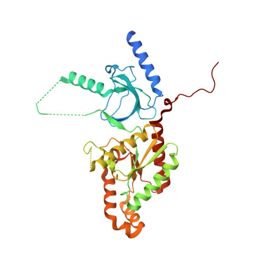
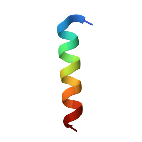
Stapled Voltage-Gated Calcium Channel (CaV) alpha-Interaction Domain (AID) Peptides Act As Selective Protein-Protein Interaction Inhibitors of CaV Function.

Findeisen, F.Campiglio, M.Jo, H.Abderemane-Ali, F.Rumpf, C.H.Pope, L.Rossen, N.D.Flucher, B.E.DeGrado, W.F.Minor, D.L.

(2017) ACS Chem Neurosci 8: 1313-1326

  • DOI: https://doi.org/10.1021/acschemneuro.6b00454
  • Primary Citation Related Structures: 
    5V2P, 5V2Q

  • PubMed Abstract: 

    For many voltage-gated ion channels (VGICs), creation of a properly functioning ion channel requires the formation of specific protein-protein interactions between the transmembrane pore-forming subunits and cystoplasmic accessory subunits. Despite the importance of such protein-protein interactions in VGIC function and assembly, their potential as sites for VGIC modulator development has been largely overlooked. Here, we develop meta-xylyl (m-xylyl) stapled peptides that target a prototypic VGIC high affinity protein-protein interaction, the interaction between the voltage-gated calcium channel (Ca V ) pore-forming subunit α-interaction domain (AID) and cytoplasmic β-subunit (Ca V β). We show using circular dichroism spectroscopy, X-ray crystallography, and isothermal titration calorimetry that the m-xylyl staples enhance AID helix formation are structurally compatible with native-like AID:Ca V β interactions and reduce the entropic penalty associated with AID binding to Ca V β. Importantly, electrophysiological studies reveal that stapled AID peptides act as effective inhibitors of the Ca V α 1 :Ca V β interaction that modulate Ca V function in an Ca V β isoform-selective manner. Together, our studies provide a proof-of-concept demonstration of the use of protein-protein interaction inhibitors to control VGIC function and point to strategies for improved AID-based Ca V modulator design.


  • Organizational Affiliation
    • Molecular Biophysics & Integrated Imaging Division, Lawrence Berkeley National Laboratory , Berkeley, California 94720, United States.

Macromolecule Content 

  • Total Structure Weight: 41.54 kDa 
  • Atom Count: 2,997 
  • Modeled Residue Count: 317 
  • Deposited Residue Count: 365 
  • Unique protein chains: 2

Macromolecules

Find similar proteins by:|  3D Structure
Entity ID: 1
MoleculeChains  Sequence LengthOrganismDetailsImage
Voltage-dependent L-type calcium channel subunit beta-2,Voltage-dependent L-type calcium channel subunit beta-2346Rattus norvegicusMutation(s): 0 
Gene Names: Cacnb2Cacnlb2
UniProt
Find proteins for Q8VGC3 (Rattus norvegicus)
Explore Q8VGC3 
Go to UniProtKB:  Q8VGC3
Entity Groups
Sequence Clusters30% Identity50% Identity70% Identity90% Identity95% Identity100% Identity
UniProt GroupQ8VGC3
Sequence Annotations
Expand
Reference Sequence
Find similar proteins by:  Sequence   |   3D Structure  
Entity ID: 2
MoleculeChains  Sequence LengthOrganismDetailsImage
Voltage-dependent L-type calcium channel subunit alpha-1C19Homo sapiensMutation(s): 5 
UniProt & NIH Common Fund Data Resources
Find proteins for Q13936 (Homo sapiens)
Explore Q13936 
Go to UniProtKB:  Q13936
PHAROS:  Q13936
GTEx:  ENSG00000151067 
Entity Groups
Sequence Clusters30% Identity50% Identity70% Identity90% Identity95% Identity100% Identity
UniProt GroupQ13936
Sequence Annotations
Expand
Reference Sequence

Experimental Data & Validation

Experimental Data

  • Method: X-RAY DIFFRACTION
  • Resolution: 1.70 Å
  • R-Value Free:  0.196 (Depositor), 0.203 (DCC) 
  • R-Value Work:  0.154 (Depositor), 0.169 (DCC) 
  • R-Value Observed: 0.156 (Depositor) 
Space Group: P 21 21 21
Unit Cell:
Length ( Å )Angle ( ˚ )
a = 54.975α = 90
b = 61.92β = 90
c = 129.877γ = 90
Software Package:
Software NamePurpose
REFMACrefinement
iMOSFLMdata reduction
SCALAdata scaling
PHASERphasing

Structure Validation

View Full Validation Report


Ligand Structure Quality Assessment 


Entry History 

& Funding Information

Deposition Data


Funding OrganizationLocationGrant Number
National Institutes of Health/National Heart, Lung, and Blood Institute (NIH/NHLBI)United StatesR01-HL080050
National Institutes of Health/National Institute of General Medical Sciences (NIH/NIGMS)United StatesR01-GM54616

Revision History  (Full details and data files)

  • Version 1.0: 2017-07-19
    Type: Initial release
  • Version 1.1: 2017-08-23
    Changes: Data collection
  • Version 1.2: 2017-09-13
    Changes: Author supporting evidence
  • Version 1.3: 2019-12-04
    Changes: Author supporting evidence
  • Version 1.4: 2023-10-04
    Changes: Data collection, Database references, Refinement description
  • Version 1.5: 2024-11-20
    Changes: Structure summary