5UVG | pdb_00005uvg

Crystal structure of the human neutral sphingomyelinase 2 (nSMase2) catalytic domain with insertion deleted and calcium bound

  • Classification: HYDROLASE
  • Organism(s): Homo sapiens
  • Expression System: Escherichia coli
  • Mutation(s): No 
  • Membrane Protein: Yes  OPM

  • Deposited: 2017-02-20 Released: 2017-06-28 
  • Deposition Author(s): Airola, M.V., Guja, K.E., Garcia-Diaz, M., Hannun, Y.A.
  • Funding Organization(s): National Institutes of Health/National Institute of General Medical Sciences (NIH/NIGMS), National Institutes of Health/National Institute of Environmental Health Sciences (NIH/NIEHS)

Experimental Data Snapshot

  • Method: X-RAY DIFFRACTION
  • Resolution: 1.85 Å
  • R-Value Free: 
    0.197 (Depositor), 0.199 (DCC) 
  • R-Value Work: 
    0.163 (Depositor), 0.162 (DCC) 
  • R-Value Observed: 
    0.164 (Depositor) 

wwPDB Validation 3D Report Full Report

Validation slider image for 5UVG

This is version 1.4 of the entry. See complete history

Literature

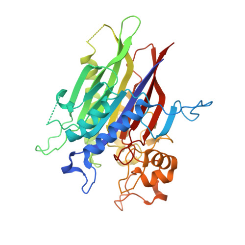
Structure of human nSMase2 reveals an interdomain allosteric activation mechanism for ceramide generation.

Airola, M.V.Shanbhogue, P.Shamseddine, A.A.Guja, K.E.Senkal, C.E.Maini, R.Bartke, N.Wu, B.X.Obeid, L.M.Garcia-Diaz, M.Hannun, Y.A.

(2017) Proc Natl Acad Sci U S A 114: E5549-E5558

  • DOI: https://doi.org/10.1073/pnas.1705134114
  • Primary Citation Related Structures: 
    5UVG

  • PubMed Abstract: 

    Neutral sphingomyelinase 2 (nSMase2, product of the SMPD3 gene) is a key enzyme for ceramide generation that is involved in regulating cellular stress responses and exosome-mediated intercellular communication. nSMase2 is activated by diverse stimuli, including the anionic phospholipid phosphatidylserine. Phosphatidylserine binds to an integral-membrane N-terminal domain (NTD); however, how the NTD activates the C-terminal catalytic domain is unclear. Here, we identify the complete catalytic domain of nSMase2, which was misannotated because of a large insertion. We find the soluble catalytic domain interacts directly with the membrane-associated NTD, which serves as both a membrane anchor and an allosteric activator. The juxtamembrane region, which links the NTD and the catalytic domain, is necessary and sufficient for activation. Furthermore, we provide a mechanistic basis for this phenomenon using the crystal structure of the human nSMase2 catalytic domain determined at 1.85-Å resolution. The structure reveals a DNase-I-type fold with a hydrophobic track leading to the active site that is blocked by an evolutionarily conserved motif which we term the "DK switch." Structural analysis of nSMase2 and the extended N-SMase family shows that the DK switch can adopt different conformations to reposition a universally conserved Asp (D) residue involved in catalysis. Mutation of this Asp residue in nSMase2 disrupts catalysis, allosteric activation, stimulation by phosphatidylserine, and pharmacological inhibition by the lipid-competitive inhibitor GW4869. Taken together, these results demonstrate that the DK switch regulates ceramide generation by nSMase2 and is governed by an allosteric interdomain interaction at the membrane interface.


  • Organizational Affiliation
    • Stony Brook University Cancer Center, Stony Brook, NY 11794.

Macromolecule Content 

  • Total Structure Weight: 41.39 kDa 
  • Atom Count: 2,966 
  • Modeled Residue Count: 348 
  • Deposited Residue Count: 371 
  • Unique protein chains: 1

Macromolecules

Find similar proteins by:|  3D Structure
Entity ID: 1
MoleculeChains  Sequence LengthOrganismDetailsImage
Sphingomyelin phosphodiesterase 3,Sphingomyelin phosphodiesterase 3371Homo sapiensMutation(s): 0 
Gene Names: SMPD3
EC: 3.1.4.12
Membrane Entity: Yes 
UniProt & NIH Common Fund Data Resources
Find proteins for Q9NY59 (Homo sapiens)
Explore Q9NY59 
Go to UniProtKB:  Q9NY59
PHAROS:  Q9NY59
GTEx:  ENSG00000103056 
Entity Groups
Sequence Clusters30% Identity50% Identity70% Identity90% Identity95% Identity100% Identity
UniProt GroupQ9NY59
Sequence Annotations
Expand
Reference Sequence

Small Molecules

Ligands 1 Unique
IDChains Name / Formula / InChI Key2D Diagram3D Interactions
CA

Query on CA


Download:Ideal Coordinates CCD File
B [auth A]CALCIUM ION
Ca
BHPQYMZQTOCNFJ-UHFFFAOYSA-N

Experimental Data & Validation

Experimental Data

  • Method: X-RAY DIFFRACTION
  • Resolution: 1.85 Å
  • R-Value Free:  0.197 (Depositor), 0.199 (DCC) 
  • R-Value Work:  0.163 (Depositor), 0.162 (DCC) 
  • R-Value Observed: 0.164 (Depositor) 
Space Group: P 21 21 2
Unit Cell:
Length ( Å )Angle ( ˚ )
a = 73.61α = 90
b = 91.05β = 90
c = 50.03γ = 90
Software Package:
Software NamePurpose
Aimlessdata scaling
BUSTERrefinement
PDB_EXTRACTdata extraction
XDSdata reduction
SHARPphasing

Structure Validation

View Full Validation Report


Entry History 

& Funding Information

Deposition Data


Funding OrganizationLocationGrant Number
National Institutes of Health/National Institute of General Medical Sciences (NIH/NIGMS)United StatesR37GM043825
National Institutes of Health/National Institute of General Medical Sciences (NIH/NIGMS)United StatesF32GM100679
National Institutes of Health/National Institute of General Medical Sciences (NIH/NIGMS)United StatesR01GM100021
National Institutes of Health/National Institute of Environmental Health Sciences (NIH/NIEHS)United StatesF30ES022930

Revision History  (Full details and data files)

  • Version 1.0: 2017-06-28
    Type: Initial release
  • Version 1.1: 2017-09-20
    Changes: Author supporting evidence
  • Version 1.2: 2018-05-09
    Changes: Data collection, Database references
  • Version 1.3: 2019-12-18
    Changes: Author supporting evidence
  • Version 1.4: 2024-03-06
    Changes: Data collection, Database references, Derived calculations